Quantitative Iris Phenotype–Response Coupling During Selective Stromal Melanin Modulation: A Prospective Longitudinal Research Framework
Complete source PDF remains authoritative for embedded figures, formulas and tabular layout. The web reading edition reproduces the conceptual research text, not an independent validation study.
Article type: Research Concept / Prospective Validation Framework Version: 1.2 Date: October 2026 Reserved Zenodo DOI: 10.5281/zenodo.23142713 Evidence status: Conceptual framework; no prospective patient-level validation dataset Prepared for Zenodo deposition Suggested citation: Mete M. Quantitative Iris Phenotype–Response Coupling During Selective Stromal Melanin Modulation: A Prospective Longitudinal Research Framework. Version 1.2. Zenodo; 2026. doi:10.5281/zenodo.23142713.
Abstract
Background Human iris appearance is the optical expression of a multidimensional tissue phenotype involving stromal pigmentation, tissue architecture, light scattering, collagen organization, vascular characteristics, pupil state, and spatial variation in pigment distribution. Conventional descriptors such as brown, hazel, green, grey, or blue therefore provide only a limited representation of the underlying phenotype. Standardized photography and quantitative colorimetry permit continuous measurement of visible iris characteristics, while anterior-segment optical coherence tomography (AS-OCT) enables structural characterization of iris geometry and regional thickness.[1–3] More recently, polarization-sensitive AS-OCT has demonstrated the feasibility of deriving melanin-related quantitative measurements from the iris stroma.[4]
Objective This research concept proposes a prospective longitudinal framework for examining whether measurable pretreatment iris phenotype is associated with the subsequent trajectory of response during response-guided selective stromal melanin modulation.
The principal objective is not prediction of a predetermined final eye color. Instead, the framework investigates whether baseline tissue characteristics are associated with dynamic response properties including response slope, cumulative treatment exposure, spatial heterogeneity, delayed post-exposure evolution, and response plateau.
Central Hypothesis The central hypothesis is that longitudinal response is not determined by apparent iris color alone. Rather, the observed trajectory of stromal pigment modification reflects the interaction of baseline pigment burden, iris architecture, spatial heterogeneity, optical characteristics, cumulative treatment exposure, and time-dependent biological processing.
Proposed Approach Pretreatment characterization would combine standardized iris photography, quantitative colorimetry, clinically applicable pigmentation grading, structural AS-OCT, and technically qualified OCT-derived optical variables.
These baseline variables would be related prospectively to longitudinal outcomes including: • early quantitative response slope; • exposure-normalized change; • cumulative documented exposure burden across longitudinal observation; • sectoral response heterogeneity; • delayed change following cessation of active treatment exposure; and • longitudinal plateau-like behavior.
Longitudinal hierarchical modeling would account for repeated observations, bilateral-eye dependence, participant-level variability, cumulative treatment exposure, and potentially nonlinear trajectories.
Significance The proposed framework shifts investigation from static color classification toward quantitative phenotype– response coupling. If validated, it could clarify why irides with superficially similar baseline appearance may exhibit different longitudinal responses.
This framework is hypothesis-generating. It does not constitute a validated treatment algorithm, safety algorithm, final-color prediction model, treatment-prescription system, or guarantee of a specific cosmetic outcome.
Keywords: iris pigmentation; quantitative iris phenotyping; anterior-segment OCT; selective stromal melanin modulation; laser
iris depigmentation; longitudinal response; response kinetics; iris stroma; quantitative colorimetry; spatial heterogeneity; plateau dynamics.
1. Introduction
Human iris color is usually communicated through a small number of categorical labels. However, visible color represents the macroscopic result of several interacting biological and optical variables rather than a single pigment measurement.
Quantitative photographic studies have demonstrated that iris pigmentation can be represented continuously using standardized colorimetric variables rather than being restricted to subjective color categories.[1]
Structural imaging studies have independently demonstrated measurable regional variation in iris thickness, volume, width, and morphology using anterior-segment OCT.[2,3]
These observations support a more quantitative representation of the iris phenotype.
More recently, Li et al. used polarization-sensitive anterior-segment OCT to derive an iris stromal melanin concentration ratio and demonstrated significant relationships between this measurement and CIE L*a*b* characteristics of visible iris color.[4] This work further supports the principle that aspects of iris pigmentation can be investigated as measurable tissue characteristics rather than solely as externally perceived color.
A different scientific question emerges when anterior stromal pigmentation is intentionally modified.
Experimental iris-laser work and published human clinical series demonstrate that visible pigment change may evolve over time rather than representing an instantaneous fixed endpoint.[5,6] However, the existing literature does not establish that all procedures grouped under the broad term laser iris depigmentation (LID) represent an equivalent intervention. Published reports and reviews encompass differing treatment concepts, exposure architectures, devices, treatment schedules, treated areas, and clinical management strategies.[6–8]
Accordingly, procedural labels alone should not be assumed to establish biological or clinical equivalence.
Within the present research program, Lumineyes™ is considered separately as a response-guided staged approach based on selective stromal melanin modulation.[13,14] The broader LID literature is therefore used as contextual evidence rather than being treated as directly interchangeable evidence for Lumineyes™.
The unresolved scientific question addressed here is: Does the quantitative pretreatment iris phenotype contain information about the trajectory by which that iris subsequently responds during response-guided selective stromal melanin modulation?
This question forms the basis of the proposed phenotype–response framework.
2. Terminological and Procedural Boundary
The phrase laser iris depigmentation describes a broad procedural category in the published literature rather than a single universally standardized intervention.[6–8]
This distinction is methodologically important.
A complication, treatment response, exposure characteristic, or temporal pattern observed with one published LID technique should not automatically be attributed to another technique unless sufficient procedural equivalence has been demonstrated.
Likewise, findings obtained within the Lumineyes™ framework should not automatically be generalized to unrelated LID procedures.
For the purposes of the present framework: • Laser iris depigmentation (LID) refers primarily to the heterogeneous broader literature concerning laser-based reduction of visible anterior iris pigmentation. • Selective stromal melanin modulation (SSMM) refers to the treatment-target concept used within the Lumineyes™ research framework. • Lumineyes™ refers to a response-guided staged clinical framework in which sequential exposure is considered in relation to observed tissue response rather than being represented simply by an invariant predetermined session sequence.[13,14]
This terminology is intended to improve methodological specificity rather than imply comparative superiority.
3. Scientific Scope
The present framework separates four related but analytically distinct domains.
3.1. Baseline phenotype
What measurable structural, pigmentary, spatial, and optical characteristics describe the untreated iris?
3.2. Treatment exposure
What cumulative controlled exposure has occurred over time?
3.3. Biological and optical response
How does the measurable iris phenotype change following exposure?
3.4. Longitudinal kinetics
At what rate, spatial distribution, and temporal pattern does that response evolve?
The present concept is concerned primarily with the coupling between baseline phenotype and longitudinal response kinetics.
It is not designed to predict a predetermined cosmetic final color.
Earlier patent literature has already described approaches intended to predict or simulate post-treatment perceived iris color using anatomical, phenotypic, photographic, or other patient-specific information.[11,12]
The present framework addresses a different endpoint.
Rather than asking: What exact color will this eye become?
It asks: How does a quantitatively characterized iris change as a function of controlled exposure and time?
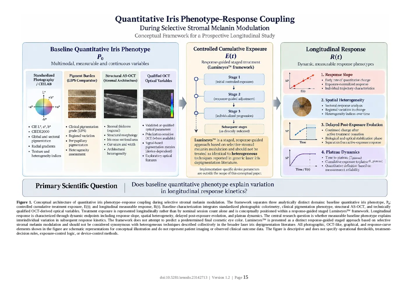
The principal dynamic endpoints are therefore: • response slope; • exposure-normalized response; • cumulative exposure burden; • spatial response heterogeneity; • delayed post-exposure evolution; and • plateau dynamics.
4. Relationship to Existing MyLumineyes Research Frameworks
The proposed work complements rather than duplicates existing research concepts.
4.1. Lumineyes Optical Architectural Typology
The Lumineyes Optical Architectural Typology (L-OAT) proposes a multimodal framework for describing baseline iris phenotype using visible appearance, architecture, and qualified OCT-derived signal characteristics. [13]
Its primary question is: What is the measurable phenotype of the iris at baseline?
The present framework begins where that question ends: Does the baseline phenotype contain information about subsequent response behavior?
Accordingly: L-OAT = baseline phenotype characterization whereas the present framework addresses: baseline phenotype → longitudinal response coupling
4.2. Clinical Pigmentation Grading
An ordinal clinical pigmentation descriptor, including LIPS where used within the Lumineyes™ research program, may remain useful as a pragmatic baseline comparator.
However, ordinal pigmentation grade should not be assumed to capture all relevant biological variation.
A principal empirical question of the proposed study is therefore whether continuous multimodal phenotype measurements provide information beyond a relatively simple clinical pigmentation grade.
4.3. Temporal Response Architecture
Temporal Response Architecture (TRA) organizes potential post-exposure anterior-segment responses according to interacting temporal domains and explicitly distinguishes elapsed time from biological state and longitudinal trajectory.[14]
TRA asks: What response processes may evolve over time after exposure?
The present framework asks: Can characteristics measured before exposure explain variation in those subsequent trajectories?
The two frameworks are therefore complementary: TRA: exposure → temporal response biology Present framework: baseline phenotype → variation in temporal response Neither framework should be treated as validation of the other.
5. Central Hypothesis
The central hypothesis is: Longitudinal response during selective stromal melanin modulation is partly associated with measurable pretreatment iris phenotype, and a multidimensional baseline phenotype will explain response variability more effectively than conventional visible-color classification alone.
This proposition generates several testable subhypotheses.
H1 – Baseline pigment burden Greater quantitative baseline stromal pigmentation may be associated with a different response slope and greater cumulative exposure burden across the observed response trajectory.
H2 – Stromal architecture Reproducible structural characteristics may contribute information about longitudinal response independently of apparent baseline color.
H3 – Spatial heterogeneity Greater pretreatment regional pigmentation or architectural heterogeneity may be associated with greater spatial variation in subsequent response.
H4 – Multimodal phenotype A model incorporating standardized photographic phenotype, structural architecture, and technically qualified OCT variables may explain longitudinal response more effectively than categorical color classification alone.
H5 – Nonlinear kinetics Response may not remain linear across staged treatment. Individual trajectories may demonstrate rapid early change, progressive slowing, delayed post-exposure evolution, or plateau-like behavior.
6. Conceptual Architecture
The proposed analytical structure contains three principal components: P₀ → E(t) → R(t) where: P₀ = baseline quantitative iris phenotype; E(t) = cumulative controlled treatment exposure over time; R(t) = longitudinal measurable response.
The principal scientific relationship may therefore be represented conceptually as: R(t) = f[P₀, E(t), t, participant-level factors]
This expression is a conceptual research model and not a validated biological equation.
The framework does not assume that baseline phenotype alone determines response. Rather, it tests whether baseline characteristics explain a reproducible proportion of the observed variability after treatment exposure and time are taken into account.
7. Baseline Quantitative Phenotype
Baseline phenotype should be represented using multimodal measurements while preserving a strong distinction between validated measurements, qualified surrogate variables, and exploratory features.
7.1. Standardized visible-light photography
High-resolution iris photography should be acquired under controlled conditions including: • illumination; • camera geometry; • magnification; • focus; • white balance; • pupil state; • image-processing pipeline.
Candidate quantitative variables include: • CIE L*; • CIE a*; • CIE b*; • CIEDE2000 color difference; • global pigmentation intensity; • sectoral pigmentation; • radial pigment gradients; • peripupillary pigmentation; • circumferential variation; • image-derived texture; • spatial heterogeneity indices.
The feasibility of quantitative iris-color measurement from standardized photography is established.[1]
Regional measurements should be favored where technically reproducible because a single global mean may obscure spatially heterogeneous pigmentation.
8. Structural Iris Phenotype
AS-OCT provides complementary anatomical information not contained in visible photography.
Studies have demonstrated that iris thickness and three-dimensional morphometric characteristics can be measured in vivo using OCT-based approaches.[2,3]
Candidate structural variables may include: • stromal thickness at standardized anatomical positions; • regional thickness variation; • iris cross-sectional area; • iris curvature; • pupil diameter; • regional architectural heterogeneity; • other reproducible anterior-segment anatomical covariates.
Three-dimensional variables should only be used when the acquisition protocol genuinely supports volumetric measurement.
Candidate parameters should not be retained simply because they are technically obtainable. Measurement repeatability should be established before their inclusion in predictive modeling.
9. OCT-Derived Optical Variables
OCT signal characteristics require greater interpretive caution than geometric measurements.
Conventional AS-OCT display brightness should not automatically be interpreted as direct melanin concentration.
Measured signal may depend on: • imaging wavelength; • focus; • scan geometry; • tissue orientation; • detector sensitivity; • sensitivity roll-off; • signal floor; • segmentation; • averaging; • gain; • reconstruction; • post-processing; • device-specific export behavior.
Accordingly, candidate OCT variables should be classified according to evidentiary status.
9.1. Validated or physically interpretable variables
Measurements for which an appropriate physical or experimental interpretation has been established.
9.2. Qualified surrogate variables
Repeatable signal features that may be associated with tissue characteristics but should not be represented as direct melanin measurements.
9.3. Exploratory optical variables
Candidate features requiring technical and biological validation before clinical interpretation.
Polarization-sensitive AS-OCT represents a distinct measurement approach. Li et al. demonstrated a PS-AS-OCT-derived stromal melanin concentration ratio associated with visible iris color, providing evidence that melanin-related quantitative optical assessment is feasible with appropriately specialized instrumentation.[4]
That finding should not be extrapolated to ordinary intensity-only AS-OCT systems.
10. Treatment Exposure as an Independent Variable
Treatment exposure and biological response must be analytically separated.
Repeated treatment should not simply be represented as: session 1 → session 2 → session 3 because nominal session count does not necessarily represent equivalent tissue exposure.
The prospective study should therefore represent treatment exposure longitudinally using a prespecified, internally consistent descriptor, denoted conceptually as: E(t) The descriptor is intended only to support longitudinal research analysis and comparison.
Its exact construction, weighting, parameterization, and operational implementation are outside the scope of this conceptual publication.
Implementation-specific engineering parameters, operational thresholds, treatment-decision rules, exposure-control logic, software architecture, and device-control methods are outside the scope of this conceptual publication.
The scientific requirement is sufficiently consistent internal exposure documentation to permit valid longitudinal analysis without specifying a treatment-prescription or control algorithm.
11. Longitudinal Response Phenotype
The central outcome is not a target color.
It is the trajectory of measurable change.
11.1. Response slope
The rate of quantitative phenotype change may be characterized longitudinally in relation to time and documented exposure.
In this framework, response slope is a research descriptor rather than a treatment-decision rule.
11.2. Exposure-normalized response
Individuals may differ in the magnitude of measurable change observed for comparable cumulative exposure.
An exposure-normalized response parameter may therefore provide more meaningful information than treatment-session count alone.
11.3. Cumulative exposure burden
Cumulative documented exposure may be retained as a longitudinal covariate and research outcome when comparing response trajectories.
This concept does not specify a treatment threshold, dosing rule, retreatment criterion, or action trigger.
11.4. Spatial response heterogeneity
The iris should not automatically be treated as a spatially uniform surface.
Sectoral measurements permit longitudinal estimation of regional response variation.
Spatial heterogeneity should be evaluated using prespecified sectoral analyses appropriate to the measurement platform.
No specific heterogeneity algorithm, weighting scheme, or operational threshold is prescribed in this conceptual framework.
11.5. Delayed post-exposure evolution
Visible phenotype may continue to evolve after active treatment exposure has ceased.
Accordingly, the model distinguishes: active-exposure response from post-exposure evolution.
This distinction is consistent with the broader principle that elapsed time, immediate appearance, and biological trajectory should not be treated as interchangeable constructs.[14]
12. Plateau Dynamics
Response plateau represents a potentially important longitudinal phenotype.
Plateau-like behavior should be treated as a longitudinal research phenotype rather than an informal clinical impression.
Future validation studies may investigate whether response trajectories demonstrate reproducible stabilization over time.
The present conceptual paper intentionally does not specify an operational plateau threshold, exposure trigger, stopping rule, or retreatment rule.
Its scientific objective is narrower: Does plateau behavior differ systematically according to baseline phenotype?
13. Thermal Relaxation Time Is Not Clinical Response Time
Physical laser-tissue interaction and longitudinal clinical response operate on fundamentally different timescales.
Thermal relaxation time depends on physical properties including target dimensions and thermal diffusivity.
It cannot be validly inferred solely from visible iris color, pigmentation grade, or stromal thickness.
The present framework therefore does not attempt to derive tissue thermal relaxation time from clinical imaging.
Instead, it investigates response kinetics across clinically observable intervals.
The relevant question is: How does the measurable tissue phenotype evolve following controlled exposure?
This distinction avoids conflating short-timescale laser physics with longer-timescale tissue and optical evolution.
14. Proposed Prospective Cohort
A future empirical validation study could use a prospective longitudinal observational cohort of individuals undergoing clinically determined response-guided selective stromal melanin modulation.
Research participation should not require additional treatment exposure solely to satisfy the predictive model.
Clinical treatment and safety decisions should remain governed independently from the observational research protocol.
An initial development cohort of approximately 50–100 treated eyes may be considered for feasibility planning.
However, final sample size should be determined using formal power or precision calculations informed by pilot measurement variance, within-participant correlation, anticipated effect size, repeated observations, and model complexity.
Both eyes from one participant must not be treated as independent participants.
15. Proposed Assessment Schedule
A standardized longitudinal acquisition structure may include: T₀ – Baseline Complete multimodal phenotype acquisition before active treatment.
Sequential staged assessments Standardized photography and selected clinical or imaging measurements before subsequent exposure whenever clinically feasible.
Early post-exposure assessment Characterization of immediate or early response.
Approximately 1 month Early stabilization assessment.
Approximately 3 months Intermediate response assessment.
Approximately 6 months Late response and provisional plateau assessment.
Longer follow-up should be incorporated where available.
The exact acquisition schedule should be predefined before prospective enrollment.
Time should additionally be retained as a continuous variable rather than analyzed exclusively through categorical visit labels.
16. Primary Outcomes
Two complementary primary outcomes are proposed.
Primary Outcome 1 Longitudinal quantitative response slope This represents the rate of change in a prespecified phenotype measurement as a function of time and cumulative exposure.
Primary Outcome 2 Cumulative documented exposure burden as a longitudinal research variable This characterizes exposure burden without assuming a fixed number of treatment stages or specifying a treatment threshold.
These outcomes characterize different aspects of response and should not automatically be collapsed into one composite score.
17. Secondary Outcomes
Potential secondary outcomes include: • ΔL*; • Δa*; • Δb*; • CIEDE2000 change; • regional pigmentation variance; • sectoral response concordance; • delayed post-exposure change; • time to plateau; • longitudinal plateau-like behavior; • quantitative pigmentation-index change; • technically qualified OCT-signal change; • inter-eye response concordance; • association between clinical pigmentation grade and continuous response variables; • incremental information contributed by L-OAT-derived variables beyond visible pigmentation alone.
Safety outcomes should be recorded prospectively but analyzed independently from cosmetic or pigment-response endpoints.
18. Statistical Architecture
The proposed study should prioritize longitudinal hierarchical modeling rather than isolated before-and-after comparisons.
A conceptual mixed-effects formulation may be represented as: R = β₀ + β₁ P + β₂ E + β₃ T + β₄ (P × E ) + u + v + ε ijt ij0 ijt t ij0 ijt i ij ijt where: i = participant; j = eye; t = observation time; P₀ = baseline phenotype; E = cumulative treatment exposure; u = participant-level random effect; i vij = eye-level effect nested within participant; ε = residual error. ijt The exact functional form should be data-driven only within prespecified analytical boundaries.
Nonlinear terms, restricted splines, or asymptotic functions may be considered if response trajectories demonstrate reproducible nonlinearity or plateau behavior.
19. Hierarchical Model Comparison
The incremental value of quantitative phenotyping should be tested rather than presumed.
A staged model hierarchy is proposed.
Model A – Conventional clinical phenotype Visible pigmentation category or clinical pigmentation grade.
Model B – Quantitative photographic phenotype Standardized continuous colorimetric and spatial photographic variables.
Model C – Photography plus structural phenotype Quantitative photography combined with reproducible structural AS-OCT variables.
Model D – Multimodal phenotype Photography, structural AS-OCT, and technically qualified optical variables.
Additional model complexity should only be considered valuable if it produces reproducible incremental information.
20. Validation Strategy
Multimodal longitudinal studies are particularly vulnerable to overfitting and data leakage.
Accordingly: • both eyes from the same participant must remain within the same dataset partition; • serial images from one participant must not be separated across training and validation datasets; • preprocessing and feature selection must occur within resampling loops; • model complexity must remain proportional to effective sample size; • missing-data mechanisms should be examined explicitly; • calibration should be assessed in addition to conventional performance metrics.
Internal validation may use participant-level bootstrapping or cross-validation.
Definitive generalizability would require a temporally or geographically independent validation cohort.
21. Continuous Phenotypes Before Artificial Biotypes
The term iris biotype may be clinically useful, but discrete biological classes should not be assumed before empirical evidence exists.
Primary analysis should therefore preserve continuous variables wherever possible.
Potential clusters may later emerge from combinations of: • pigment burden; • stromal thickness; • architecture; • spatial heterogeneity; • qualified optical characteristics.
Any resulting cluster should initially be treated as exploratory.
Only reproducible clusters replicated in independent populations should subsequently be interpreted as stable iris biotypes.
22. Distinction From Final-Color Prediction
The framework maintains a strict boundary between longitudinal-response characterization and aesthetic color prediction.
It does not attempt to answer: What exact named eye color will this patient ultimately obtain?
Post-treatment perceived color may be affected by residual pigmentation, stromal architecture, light scattering, pupil diameter, illumination, imaging conditions, corneal optics, and observer-dependent perception.
Previous patent literature has already described approaches intended to predict or simulate post-treatment iris color.[11,12]
The present framework instead asks: Given a quantitatively characterized baseline iris and measured cumulative exposure, what longitudinal response trajectory subsequently occurs?
A response trajectory can be scientifically measured even when the eventual perceived color cannot be predicted with certainty.
23. Safety as a Parallel but Distinct Domain
The present framework concerns phenotype–response coupling and is not itself a safety-staging algorithm.
Clinical safety variables should nevertheless be collected prospectively.
Potential domains include: • intraocular pressure; • anterior-chamber inflammatory findings; • corneal findings; • visual function; • pupil abnormalities; • patient-reported symptoms; • other prospectively defined adverse events.
Published case reports have documented severe anterior-segment complications following some laser-based cosmetic iris treatments, including marked intraocular-pressure elevation and pigmentary glaucoma.[9,10]
These reports establish that serious complications can occur within the broader field, but they should not be interpreted as establishing a uniform complication profile for every laser iris technique.
The procedural context is essential.
The published reports arose from treatment exposures whose equivalence to the Lumineyes™ response-guided staged selective stromal melanin modulation framework has not been established.[9,10,13,14]
Accordingly: Adverse events reported following other LID techniques should not automatically be attributed to Lumineyes™ without direct evidence, and observations obtained within Lumineyes™ should not automatically be generalized to unrelated LID procedures.
The appropriate scientific approach is technique-specific.
Mechanism, exposure architecture, staging, treatment response, follow-up, and safety outcome should be interpreted within the procedural framework in which the observations were generated.
Safety should therefore be analyzed in parallel with, rather than mathematically absorbed into, the phenotype– response model.
24. Scientific Novelty
The novelty of this framework should be defined narrowly.
The following concepts are not claimed as novel: • quantitative photographic assessment of iris pigmentation;[1] • CIE-based iris colorimetry;[1] • OCT measurement of iris structure and thickness;[2,3] • melanin-related assessment using polarization-sensitive AS-OCT;[4] • experimental or clinical laser modification of iris pigmentation;[5,6] • prediction or simulation of a postoperative perceived iris color.[11,12]
The proposed contribution is their organization around a different prospective research question: Are measurable pretreatment iris characteristics associated with the kinetics and spatial organization of subsequent response during response-guided selective stromal melanin modulation?
The specific longitudinal response domains proposed are:
1. quantitative response slope;
2. exposure-normalized response;
3. cumulative exposure burden in relation to longitudinal response;
4. spatial response heterogeneity;
5. delayed post-exposure evolution; and
6. longitudinal plateau behavior.
A targeted literature review performed during development of this framework did not identify a peer-reviewed prospective human study validating this complete multimodal baseline phenotype → cumulative exposure → longitudinal response kinetics architecture.
This statement should remain a provisional knowledge-gap conclusion until a formal systematic literature review has been completed.
25. Scientific Value of a Negative Result
The framework remains scientifically informative even if strong phenotype–response prediction is not demonstrated.
Failure of baseline multimodal imaging to explain meaningful response variability could indicate that: • static pretreatment phenotype has limited prognostic value; • dynamic biological processes dominate treatment response; • simpler clinical phenotyping performs comparably to complex imaging; • some candidate imaging variables are technically reproducible but biologically uninformative; • additional imaging complexity does not justify its methodological burden.
Conversely, reproducible phenotype–response coupling would justify subsequent external validation and simplification toward the smallest informative feature set.
The framework therefore evaluates the utility of quantitative phenotyping rather than assuming it in advance.
26. Limitations
This framework is conceptual and currently contains no prospective validation dataset.
Several limitations should be anticipated.
First, visible iris color is sensitive to illumination, camera characteristics, pupil state, image processing, acquisition geometry, and corneal optics.
Second, conventional OCT signal variables are device dependent and should not be interpreted automatically as direct histological or melanin measurements.
Third, treatment exposure is response-guided rather than randomly assigned. This creates potential time-dependent confounding and indication-related bias.
Fourth, bilateral eyes introduce hierarchical dependence.
Fifth, the relationship between melanin concentration, stromal architecture, measured optical signal, visible phenotype, and perceived color is likely nonlinear.
Sixth, post-exposure biological and optical evolution may continue after active treatment ceases.
Seventh, a single-center development cohort may not generalize to different populations, imaging platforms, or treatment architectures.
Eighth, association between a baseline characteristic and subsequent response does not demonstrate that the characteristic causally determines that response.
Finally, findings obtained within Lumineyes™ should not be assumed to represent the broader heterogeneous LID literature unless direct comparative evidence supports such generalization.
27. Ethical and Evidence Status
This publication describes a prospective research concept.
No patient-level prospective dataset is analyzed in this version.
Accordingly, the present manuscript does not establish: • treatment efficacy; • treatment safety; • patient-specific prognosis; • treatment eligibility; • optimal treatment exposure; • validated retreatment timing; • a validated stopping rule; • or a predictable final cosmetic result.
Future prospective research involving human participants should undergo appropriate institutional ethical review and informed-consent procedures before enrollment.
Any future predictive model should remain investigational until independently validated.
28. Conclusion
Quantitative imaging increasingly permits the iris to be described beyond conventional color labels.
Standardized photography permits continuous characterization of visible phenotype.[1] AS-OCT permits structural characterization of iris anatomy.[2,3] Advanced polarization-sensitive imaging has demonstrated the feasibility of deriving melanin-related stromal measurements.[4]
The next scientific question is not merely whether these characteristics can be measured.
It is whether they have longitudinal meaning.
The present framework proposes a prospective architecture linking: baseline quantitative iris phenotype to controlled cumulative exposure and subsequently to measurable longitudinal response.
Its central proposition is: The untreated iris may contain measurable information about the trajectory by which its phenotype subsequently evolves, even when a predetermined final perceived color cannot be reliably specified.
Testing this proposition requires longitudinal quantitative imaging, explicit exposure documentation, hierarchical statistical modeling, participant-level validation, and strict separation of static phenotype from dynamic response.
It also requires procedural specificity.
The heterogeneous broader literature of laser iris depigmentation should not be assumed to represent a single standardized intervention, and findings from one treatment architecture should not automatically be generalized to another.[6–10]
Within this research framework, Lumineyes™ is therefore evaluated specifically as a response-guided staged approach based on selective stromal melanin modulation, rather than being treated as synonymous with generic LID.[13,14]
Positive results would support quantitative phenotype–response coupling as a meaningful biological research domain; negative results would define limits on the prognostic interpretation of pretreatment iris imaging.
Figure 1. Conceptual architecture of quantitative iris phenotype–response coupling during selective stromal melanin modulation. The framework separates three analytically distinct domains: baseline quantitative iris phenotype, P₀; controlled cumulative treatment exposure, E(t); and longitudinal measurable response, R(t). Baseline characterization integrates standardized photographic colorimetry, clinical pigmentation phenotype, structural AS-OCT, and technically qualified OCT-derived optical variables. Treatment exposure is represented longitudinally rather than by nominal session count alone and is conceptually positioned within a response-guided staged Lumineyes™ framework. Longitudinal response is characterized through dynamic endpoints including response slope, spatial heterogeneity, delayed post-exposure evolution, and plateau dynamics. The central research question is whether measurable baseline phenotype explains interindividual variation in subsequent response kinetics. The framework does not attempt to predict a predetermined final cosmetic eye color. Lumineyes™ is presented as a distinct response-guided staged approach based on selective stromal melanin modulation and should not be considered synonymous with heterogeneous techniques described collectively in the broader laser iris depigmentation literature. All photographic, OCT-like, graphical, and response-curve elements shown in the figure are schematic representations for conceptual illustration and do not represent patient imaging or observed clinical outcome data. The figure is descriptive and does not specify operational thresholds, treatment-decision rules, exposure-control logic, or device-control methods.
Declarations
Evidence Status This manuscript presents a hypothesis-generating prospective research framework. It contains no prospective patient-level validation dataset.
Ethics No human participants, identifiable patient information, or newly generated clinical dataset are included in this conceptual publication. Ethical approval is therefore not applicable to the present version. Any future prospective clinical validation study will require appropriate ethical review and informed consent.
Data Availability No patient-level dataset was generated or analyzed for this conceptual publication.
Conflict of Interest Mustafa Mete, MD, is the developer of the Lumineyes™ methodology and the originator of the L-OAT and related MyLumineyes research frameworks. This relationship should be considered when interpreting the conceptual framework.
Intellectual and Methodological Boundary This manuscript describes the scientific architecture required to test phenotype–response coupling. It intentionally does not disclose implementation-specific device engineering, proprietary exposure parameters, operational thresholds, treatment-decision rules, exposure-control logic, software architecture, or device-control methods; these matters are outside the scope of the present conceptual publication.
References
- Edwards M, Gozdzik A, Ross K, Miles J, Parra EJ. Technical note: quantitative measures of iris color using high resolution photographs. Am J Phys Anthropol. 2012;147(1):141-149. doi:10.1002/ajpa.21637.
- Invernizzi A, Giardini P, Cigada M, Viola F, Staurenghi G. Three-dimensional morphometric analysis of the iris by swept-source anterior segment optical coherence tomography in a Caucasian population. Invest Ophthalmol Vis Sci. 2015;56(8):4796-4801. doi:10.1167/iovs.15-16483.
- Invernizzi A, Cigada M, Savoldi L, Cavuto S, Fontana L, Cimino L. In vivo analysis of the iris thickness by spectral domain optical coherence tomography. Br J Ophthalmol. 2014;98(9):1245-1249. doi:10.1136/bjophthalmol-2013-304481.
- Li X, Hu L, Yang B, Xiao Z, Yamanari M, Higashita R, Liu J. Quantitative measurement of iris melanin concentration by polarization-sensitive anterior segment optical coherence tomography. Eur J Ophthalmol. 2026;36(4):872-880. doi:10.1177/11206721251407025.
- Yildirim Y, Duzgun E, Kar T, Sonmez M, Kucukodaci Z, Ersanli D, Basoglu A. Evaluation of color-changing effect and complications after Nd:YAG laser application on iris surface. Med Sci Monit. 2016;22:107-114. doi:10.12659/MSM.895086.
- Grimaldos Ruiz P. Photoablative cosmetic iridoplasty: effective, safe, and predictable-eye color change in 1176 eyes. Int Ophthalmol. 2021;41(4):1381-1393. doi:10.1007/s10792-021-01693-5.
- D'Oria F, Abu-Mustafa SK, Alio JL. Cosmetic change of the apparent color of the eye: a review on surgical alternatives, outcomes and complications. Ophthalmol Ther. 2022;11(2):465-477. doi:10.1007/s40123-022-00458-2.
- D'Oria F, Alio JL. Surgical techniques for cosmetic eye color change: a narrative review. Ophthalmol Ther. 2025;14(8):1685-1694. doi:10.1007/s40123-025-01177-0.
- Liu J, Korban S, Moster MR, Rheaume MA, Wang Q. Bilateral severe iatrogenic pigmentary glaucoma following laser treatment for cosmetic iris color change. Am J Ophthalmol Case Rep. 2023;32:101927. doi:10.1016/j.ajoc.2023.101927.
- Jaber W, Nemet M, Waisbourd M. Laser iris depigmentation resulting in markedly elevated intraocular pressure and herpes simplex keratitis with corneal scarring: case report. Case Rep Ophthalmol. 2026;17(1):312-317. doi:10.1159/000551226.
- Stroma Medical Corporation. Method and apparatus for prediction of post-operative perceived iris color. WO2018033727A1. World Intellectual Property Organization; published February 22, 2018.
- Grimaldos Ruiz P. Method for predicting the aesthetic result of cosmetic laser iridoplasty. WO2019086741A2. World Intellectual Property Organization; published May 9, 2019.
- Mete M. Lumineyes Optical Architectural Typology (L-OAT): a multimodal hypothesis for quantitative iris phenotyping. Zenodo. Version 1.0. 2026. doi:10.5281/zenodo.23066772.
- Mete M. Temporal Response Architecture After Laser-Based Iris Pigment Modification: A Mechanistic Synthesis of Anterior-Segment Response Domains. Zenodo. 2026. doi:10.5281/zenodo.23128049.
Original figures and tables from the preprint
Each image is a facsimile of the indicated page in the original 2026 Zenodo PDF, not a newly generated scientific figure. The original PDF is authoritative.