Pigment Release-Clearance Balance in Selective Stromal Melanin Modulation: A Mechanistic Review and Proposed P-CLEAR Framework
Read the verified original Zenodo PDF (including original figures) · Original DOI: 10.5281/zenodo.23221292
The original preprint PDF is authoritative for figure placement, numbering and document formatting. New website graphics, if added, will be separately identified as conceptual web illustrations.
Not peer reviewed. This manuscript presents a conceptual mechanistic review and proposed research framework, not a validated safety index or retreatment protocol. This is the author-identified preprint version of the P-CLEAR manuscript prepared for journal consideration. It represents the same underlying work, rather than a separate study or independent replication.
Scope and relationship to related work
The focus of this manuscript is the time-dependent balance between pigment liberation and effective clearance, together with the proposed P-CLEAR recovery trajectories. Related author preprints address iris phenotyping, pre-exposure assessment, blood–aqueous-barrier response, temporal response domains, and procedure-specific evidence. Those topics provide context; the present contribution is the explicit release-clearance model and its proposed longitudinal recovery vocabulary. No new patient-level dataset or independently validated clinical thresholds are reported.
Abstract
Laser-induced modification of stromal iris pigment converts a brief optical exposure into an anterior-segment biological response that may evolve over hours, days, and longer intervals. Liberated pigment can interact with the trabecular meshwork and aqueous outflow system, while experimental pigment exposure alters trabecular phagocytosis, migration, cytoskeletal organization, and contractility. However, pigment-related pressure elevation cannot be explained by phagocytic failure alone. This narrative mechanistic review proposes a pigment release-clearance model in which the clinically relevant variable is the time-dependent balance between treatment-related pigment liberation and effective anterior-segment clearance. Effective clearance is considered dynamic rather than fixed and may vary with particulate burden, particle characteristics, trabecular cellular state, inflammation, aqueous outflow function, and time. The model provides a biological rationale for response-guided staging within Selective Stromal Melanin Modulation (SSMM), a proposed staged iris-pigment treatment concept. We further propose P-CLEAR (Pigment Clearance-Longitudinal Evaluation of Anterior-segment Recovery), a descriptive research framework for classifying rapid-resolving, intermediate-resolving, and delayed/persistent recovery trajectories. P-CLEAR is not presented as a retreatment algorithm, safety score, or validated index. Prospective longitudinal measurement of pigment burden, anterior-chamber inflammation, intraocular pressure, angle findings, and return toward individual baseline is required before quantitative thresholds or treatment intervals can be inferred.
Keywords: iris pigment; pigment clearance; trabecular meshwork; selective stromal melanin modulation;
laser iris depigmentation; intraocular pressure; pigmentary glaucoma; aqueous outflow; staged treatment; P-CLEAR
1. Introduction
Laser-based modification of iris pigmentation presents an unusual anterior-segment biological problem. The optical interaction responsible for altering pigment occurs over a comparatively short period, whereas the consequences of that interaction may continue to evolve after the exposure has ended. Pigment liberation, aqueous transport, cellular uptake, inflammatory activity, trabecular interaction, intraocular-pressure behavior, and biological recovery therefore occur on different temporal scales.
This distinction is clinically relevant because iris-derived pigment entering the anterior segment is not biologically inert. Pigment dispersion syndrome and pigmentary glaucoma provide established examples in which melanin accumulates within the trabecular meshwork, with a subset of affected eyes developing impaired aqueous outflow, ocular hypertension, and glaucomatous optic neuropathy.[1,2] Experimental pigmentary-glaucoma models further demonstrate that sustained pigment exposure can alter trabecular-meshwork cellular behavior and increase outflow resistance.[3,4,5]
At the same time, a simple model in which pigment mechanically obstructs the trabecular meshwork is insufficient. In an ex vivo pigmentary-glaucoma model, intraocular-pressure elevation occurred before measurable deterioration of trabecular phagocytosis, suggesting that early outflow dysfunction may involve cytoskeletal, contractile, extracellular-matrix, inflammatory, and other cellular mechanisms in addition to particulate processing.[4] Conversely, selective laser trabeculoplasty demonstrates that laser interaction with pigmented anterior-segment tissue can initiate monocyte recruitment, cytokine signaling, extracellular-matrix remodeling, and changes in Schlemm-canal permeability that modify aqueous outflow without requiring extensive structural destruction.[6,7]
The clinically relevant question may therefore be broader than whether pigment is released. It is whether treatment-related particulate burden is resolving, persisting, or accumulating within a dynamically responding anterior segment. This review develops a kinetic model around that question and proposes a longitudinal research framework for phenotyping post-exposure clearance and recovery.
2. Literature approach
This article is a narrative mechanistic review rather than a systematic review. Targeted searches were undertaken through 6 October 2026 using PubMed, publisher databases, and backward reference chaining.
Search concepts included combinations of iris pigment, pigment dispersion, pigmentary glaucoma, trabecular meshwork, phagocytosis, aqueous melanin, selective laser trabeculoplasty, laser flare photometry, laser peripheral iridotomy, laser iris depigmentation, cosmetic iris laser, refractive surgery complications, phakic intraocular lens, cataract endothelial cell loss, and endophthalmitis.
Priority was given to peer-reviewed human studies, mechanistic trabecular-meshwork and anterior-segment models, systematic reviews, large clinical series, and complication reports that could inform the biological model. The search was deliberately hypothesis-oriented: the objective was to synthesize evidence relevant to pigment burden, outflow response, recovery kinetics, and procedural heterogeneity rather than to estimate a pooled complication incidence.
3. Terminological boundaries: LID and SSMM
The term laser iris depigmentation (LID) currently encompasses heterogeneous procedures rather than a single standardized intervention. Published reports differ in laser source, wavelength, pulse characteristics, spot geometry, treatment density, treated surface area, number and spacing of sessions, perioperative medication, and follow-up architecture.[8,9,10,11,12,13,14,15,16] A shared cosmetic objective therefore does not establish biological or dosimetric equivalence.
Selective Stromal Melanin Modulation (SSMM) is used here to describe a staged treatment concept intended to preferentially interact with accessible anterior stromal melanin without deliberate incisional alteration of iris architecture, while permitting reassessment of the anterior-segment response between exposures. SSMM should be understood as a procedural and biological concept, not as histopathological proof of exclusive tissue selectivity or of absence of biological response in adjacent cells or structures.
This distinction is essential when interpreting published complications. A complication following one LID protocol cannot automatically establish the risk profile of every other laser protocol without demonstrating technical and biological comparability. Conversely, procedural heterogeneity does not make serious pigment-related adverse events irrelevant. Such reports remain important safety-boundary evidence. The appropriate approach is therefore neither universal attribution nor universal dismissal, but procedure-specific interpretation within a common anterior-segment biological framework.
4. Pigment liberation is not equivalent to pigment burden
Three related but distinct concepts should be separated: pigment liberation, pigment burden, and effective clearance. Pigment liberation describes treatment-related generation or mobilization of pigment-containing particulate material. Pigment burden describes the biologically relevant particulate material present within the anterior-segment environment at a particular time. Effective clearance describes the composite reduction of that burden through aqueous transport, cellular uptake and processing, redistribution, sequestration, and/or removal through outflow pathways.
These variables are not interchangeable. A laser exposure may produce an acute liberation event while the resulting pigment burden evolves for a substantially longer period. Similarly, visible pigment within the anterior chamber does not by itself define pathological outflow impairment. Primary pigment dispersion syndrome demonstrates that substantial pigment dispersion can exist without glaucoma, while other eyes develop pigment-related ocular hypertension and secondary glaucoma.[1,2]
Aqueous pigment is potentially measurable rather than purely theoretical. Kuechle et al. demonstrated reproducible quantification of aqueous melanin granules in pigment dispersion syndrome using the cell-count mode of a laser flare-cell meter.[17] Mardin et al. subsequently reported an association between aqueous melanin-granule number and maximum IOP in pigment dispersion syndrome and pigmentary glaucoma.[18]
These studies do not validate the same measurement method after SSMM, but they establish that dispersed aqueous melanin can be quantified and that its quantity may contain information not captured by a binary description of pigment presence or absence. Pigment burden should therefore be considered a kinetic variable.
5. Biological processing and the trabecular outflow system
5.1. Trabecular phagocytosis and cellular state
Trabecular-meshwork cells are actively phagocytic and contribute to the processing of pigment, cellular debris, and other particulate material encountered within the conventional aqueous outflow pathway.
Experimental data indicate that pigment exposure can modify this capacity. Wang et al. found that pigment-exposed trabecular cells demonstrated reduced phagocytosis and migration together with increased actin stress-fiber formation and cellular contraction.[5] In a related perfused anterior-segment model, sustained pigment exposure reproduced ocular hypertension, trabecular pigmentation, cytoskeletal alterations, and later deterioration in phagocytic function.[3]
The trabecular meshwork should therefore not be conceptualized as a passive filter with a fixed mechanical pore size. It is a biologically active tissue whose functional state may itself be altered by particulate exposure.
5.2. Phagocytic decline is not the entire mechanism
The temporal relationship between pigment exposure and phagocytosis is particularly important. Dang et al. demonstrated significant IOP elevation after experimental pigment exposure before measurable reduction in trabecular phagocytic capacity became evident.[4] Thus, early pressure elevation did not require established phagocytic failure.
This observation argues against a simplistic sequence of pigment accumulation, phagocytic saturation, obstruction, and IOP elevation as the sole mechanistic explanation. A more plausible model incorporates parallel and interacting pathways including altered trabecular contractility, cytoskeletal remodeling, cellular migration, extracellular-matrix regulation, aqueous-flow resistance, inflammatory signaling, and particulate processing.
5.3. Monocytes and macrophage-related responses
Evidence from selective laser trabeculoplasty reinforces the concept that cellular recruitment can participate in aqueous-outflow regulation. Alvarado et al. reported increased monocyte recruitment within the trabecular meshwork after SLT and demonstrated that monocytes or monocyte-derived factors could increase outflow facility and Schlemm-canal endothelial conductivity.[6] Contemporary reviews describe SLT as producing a predominantly biological response involving cytokine signaling, monocyte recruitment, extracellular-matrix remodeling, and altered Schlemm-canal permeability.[7]
SSMM and SLT are not equivalent procedures. Their anatomical target, intended effect, exposure geometry, and pigment source differ. SLT is relevant here for a narrower reason: it demonstrates that laser interaction with pigmented anterior-segment structures can initiate a temporally evolving cellular response involving the aqueous-outflow system. The anterior segment is therefore not merely a conduit through which pigment passively drains.
The proposed terminology is summarized in Table 1.
6. The pigment release-clearance balance
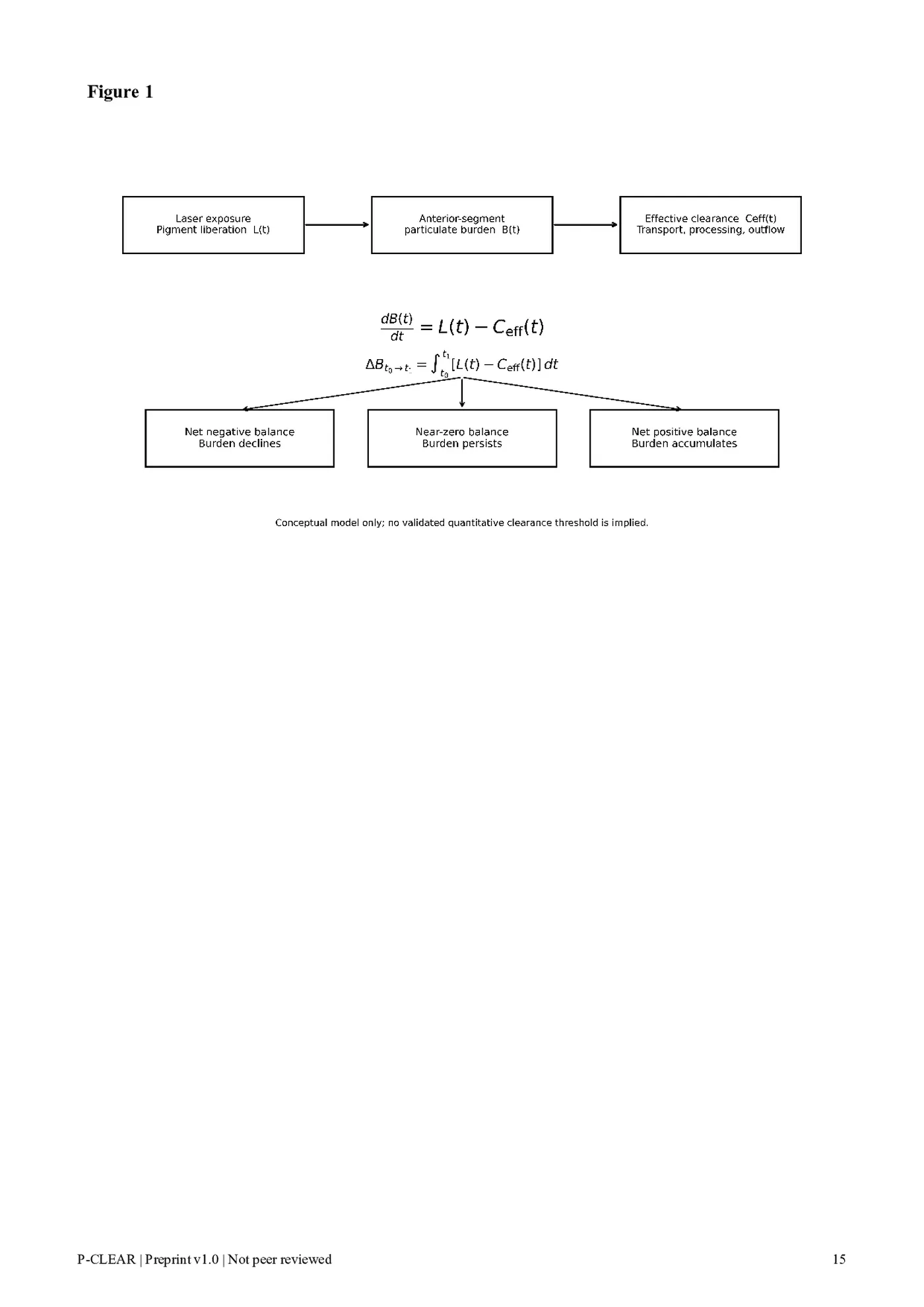
Let B(t) denote the biologically relevant anterior-segment pigment/particulate burden at time t. A simplified mass-balance representation is: dB(t)/dt = L(t) - Ceff(t) where L(t) represents treatment-related liberation or entry of pigment-associated particulate material into the anterior-segment environment and Ceff(t) represents effective biological clearance. This equation is conceptual. Neither term currently has a validated clinical unit following SSMM or other forms of laser iris pigment reduction.
An instantaneous requirement that pigment-liberation rate always remain below simultaneous clearance would be biologically unrealistic. During and immediately after exposure, liberation may transiently exceed processing. The more relevant quantity is the time-integrated balance over an observation interval: Delta B(t0 to t1) = Integral [L(t) - Ceff(t)] dt If the integrated balance becomes negative after exposure, net particulate burden moves toward resolution. If it remains approximately neutral, burden may persist. If it remains positive over a clinically meaningful interval, particulate burden accumulates. A brief liberation pulse therefore need not imply persistent accumulation if the subsequent recovery interval permits effective processing. In contrast, repeated exposure introduces a new source term before the preceding biological burden has necessarily resolved.
The central kinetic principle is therefore that potentially safety-relevant pigment kinetics depend not simply on whether liberation temporarily exceeds clearance, but on whether repeated exposure produces sustained net accumulation relative to the evolving capacity of the anterior segment to recover.
Figure 1 summarizes the release-clearance balance.
7. Clearance capacity is dynamic, not fixed
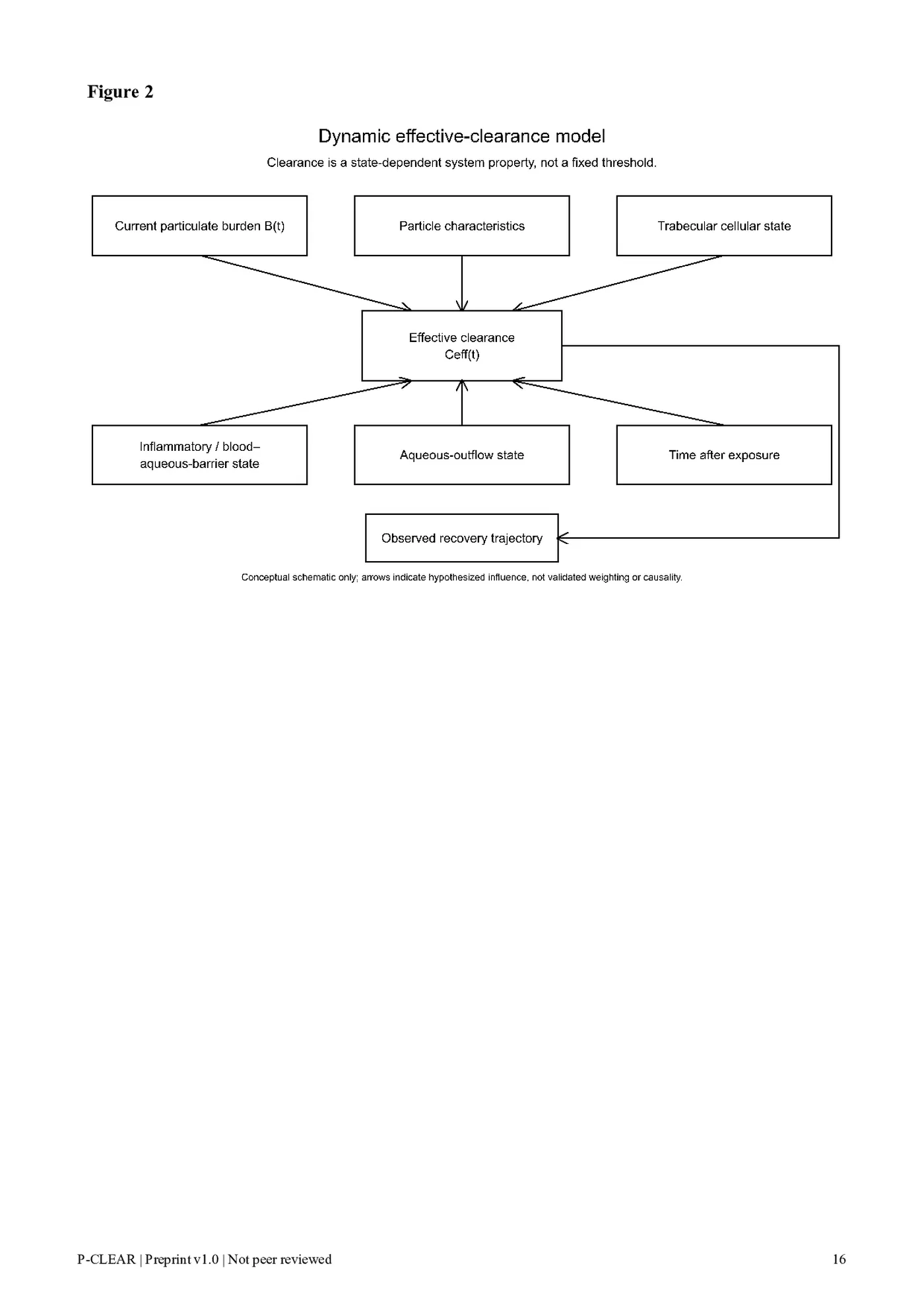
A second simplification would be to assume that effective clearance is constant. Available biology suggests otherwise. A more plausible conceptual form is: Ceff(t) = f[B(t), P, T(t), I(t), O(t), t] where B(t) represents current particulate burden; P, relevant particle characteristics; T(t), trabecular cellular state; I(t), inflammatory or blood-aqueous-barrier state; O(t), functional aqueous-outflow state; and t, time.
The expression does not imply that these variables are currently quantifiable within a validated clinical model. Its purpose is to emphasize that clearance is an emergent system property rather than a fixed numerical drainage capacity.
Experimental pigment exposure supports this interpretation. Increasing burden can alter trabecular phagocytosis, migration, contractility, and cytoskeletal organization.[3,4,5] Consequently, doubling pigment burden cannot be assumed merely to double the time required for an otherwise unchanged clearance system.
At low or transient burden, processing may remain efficient. At greater burden, recovery may become slower.
At persistent or high burden, the biological system responsible for processing and outflow may itself become altered.
This introduces the possibility of non-linearity and, under some conditions, positive feedback: greater burden may alter trabecular behavior or outflow, which may in turn slow effective recovery and prolong burden persistence. This remains a mechanistic hypothesis rather than a demonstrated threshold phenomenon after SSMM. The term trabecular saturation may therefore be intuitively useful but is biologically too narrow if interpreted literally. Clearance-capacity exceedance or persistent net particulate accumulation more accurately reflects the multi-component nature of the response.
Figure 2 summarizes the proposed determinants of effective clearance.
8. Inflammation and blood-aqueous-barrier state
Pigment kinetics should not be separated artificially from inflammation. Laser-induced anterior-segment responses may alter the blood-aqueous barrier, generate inflammatory cells and protein, and influence aqueous and trabecular physiology. Standard slit-lamp grading remains clinically important, but cells, flare, and pigment should be documented separately whenever they can be distinguished.
Laser flare photometry offers an objective method for quantifying aqueous flare and has been used to characterize inflammation following glaucoma laser and surgical procedures.[19,20] It is not interchangeable with clinical slit-lamp grading, but it can provide an additional longitudinal measurement of blood-aqueous-barrier disturbance.
The relevance to pigment clearance is twofold. First, inflammatory activity and particulate burden may occur simultaneously but follow different trajectories. Second, a clinically meaningful recovery state should not be inferred solely from disappearance of visible free pigment if flare, IOP, corneal findings, or outflow abnormalities remain active. Pigment resolution is therefore not synonymous with complete anterior-segment recovery, and persistent visible pigment does not automatically prove progressive pathological outflow failure. Longitudinal interpretation is required.
9. Why staging is a biological principle
Staged treatment is often described merely as division of a procedure into multiple sessions. From a kinetic perspective, this description is incomplete. Staging has two distinct functions. First, it distributes treatment-related pigment liberation over time rather than concentrating the intended exposure within the shortest achievable interval. Second, it creates observation windows in which the response to the preceding exposure can be evaluated before another source term is introduced.
If the anterior segment is still carrying a residual particulate, inflammatory, or outflow burden from an earlier exposure, additional treatment does not begin from baseline. It begins from an already altered state.
Accordingly, elapsed time is not equivalent to biological recovery, and a fixed interval is not itself permission for re-exposure.
The importance of time-dependent post-laser responses is well established elsewhere in ophthalmology.
Following Nd:YAG peripheral iridotomy, acute IOP elevation has been documented and related to treatment variables such as energy and pulse number.[21] The anatomy and purpose of iridotomy are entirely different from SSMM, and these data cannot define an SSMM treatment interval. They illustrate the broader principle that anterior-segment laser effects can produce measurable biological consequences after the optical event itself is complete. Staging should therefore be interpreted as a biological observation architecture, not simply a calendar.
10. Existing iris-laser literature demonstrates procedural heterogeneity
Published iris-directed laser literature reinforces the need for protocol-specific interpretation. Experimental work by Yildirim et al. evaluated 532-nm Nd:YAG laser exposure on rabbit iris and documented transient inflammatory findings with histological reduction in pigment density.[8] Basoglu and Celik subsequently described treatment of sectoral heterochromia using a frequency-doubled 532-nm SLT laser device.[9]
A substantially larger prospective series by Grimaldos Ruiz included 1176 eyes treated with a 532-nm Q-switched laser. Treatment was organized into repeated sessions and phases separated by longer intervals.
The study reported no significant pre-/post-treatment difference in IOP and described delayed, brief iritis as the principal complication.[10] These findings coexist with published reports of severe secondary pigmentary glaucoma following other cosmetic iris-laser procedures.[13,14,15,16]
The coexistence of favorable series and severe complication reports should not be resolved by treating either as universally representative. Instead, it highlights a methodological problem: laser iris depigmentation is presently an exposure category rather than a standardized dose-defined intervention. Meaningful safety reporting should therefore characterize the laser system, energy-delivery architecture, treated area, exposure density, number and spacing of sessions, cumulative exposure, medication, baseline ocular status, and longitudinal anterior-segment response. Without these variables, attribution of a biological outcome to a generic procedural name is intrinsically limited.[11,12]
11. Complication reports through a kinetic lens
Case reports of pigmentary glaucoma after cosmetic iris laser treatment establish that severe pigment-associated outflow failure is possible. Ong et al. described refractory bilateral iatrogenic pigmentary glaucoma with intense angle pigmentation requiring trabeculectomy.[13] Swampillai et al. reported secondary pigmentary glaucoma after a course of cosmetic iris laser treatment.[14] Liu et al. described a patient presenting after four bilateral sessions with IOPs of 50 and 42 mmHg, 4+ free-floating pigmented cells, and severe visual morbidity requiring urgent bilateral trabeculectomy; the laser-treatment parameters were unavailable, limiting reconstruction of the exposure mechanism.[15] Jaber et al. more recently reported markedly elevated IOP after multiple LID sessions together with additional anterior-segment morbidity.[16]
These cases establish important safety signals. They do not establish that every laser-based iris pigment-reduction protocol has the same risk. From a kinetic perspective they generate specific unanswered questions: what burden was generated by each exposure; how much remained when subsequent exposure occurred; whether the anterior chamber still showed pigment or inflammatory activity; how IOP evolved between sessions; whether angle pigmentation was changing; whether biological recovery was documented or inferred from elapsed time alone; and which treatment characteristics determined the magnitude and spatial distribution of pigment liberation. Most retrospective complication reports cannot reconstruct these variables.
Prospective studies can.
12. P-CLEAR: Pigment Clearance-Longitudinal Evaluation of Anterior-segment Recovery
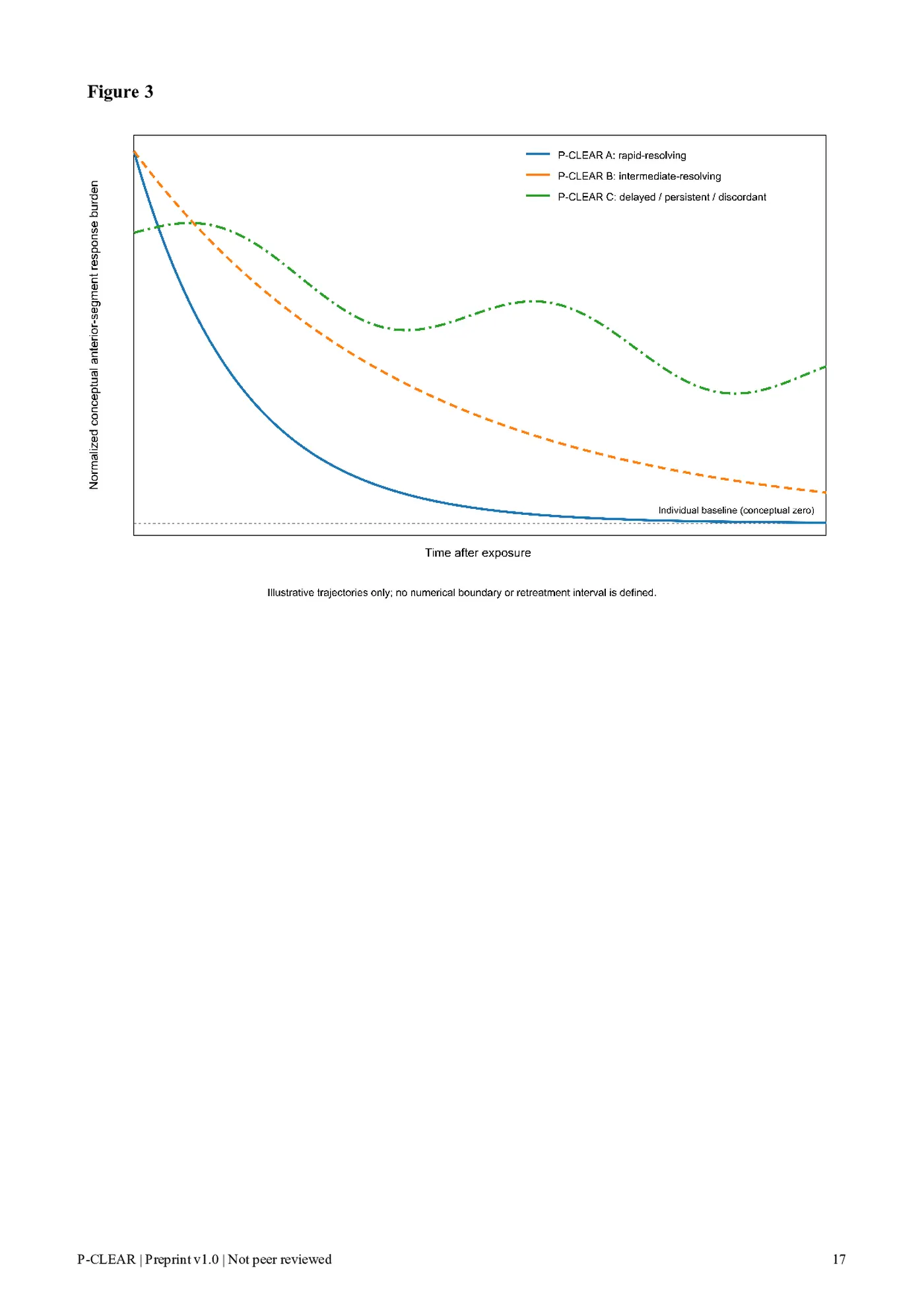
The kinetic model requires a practical observational language. We therefore propose P-CLEAR: Pigment Clearance-Longitudinal Evaluation of Anterior-segment Recovery. P-CLEAR is not a numerical index, treatment-dose calculator, complication-risk score, or automated retreatment rule. Its initial purpose is narrower: to describe candidate directions and tempos of post-exposure recovery using serial anterior-segment observations rather than elapsed time alone (Figure 3).
12.1. P-CLEAR A - rapid-resolving trajectory
A P-CLEAR A pattern would be characterized by a relatively prompt and consistent trajectory toward the eye's individual baseline, potentially including progressive decline in free pigment or particulate activity, resolving inflammatory activity, stable IOP trajectory, absence of a progressive angle/outflow signal, and recovery of other treatment-related anterior-segment findings. The defining feature is direction and consistency of recovery, not a predetermined day number.
12.2. P-CLEAR B - intermediate-resolving trajectory
A P-CLEAR B pattern would describe continuing but slower biological resolution. Potential characteristics include persistent but decreasing particulate activity, slower reduction in flare or cells, stable or recovering IOP, absence of progressively worsening outflow dysfunction, and delayed return toward individual baseline.
P-CLEAR B describes ongoing recovery, not treatment failure.
12.3. P-CLEAR C - delayed, persistent, or discordant trajectory
A P-CLEAR C pattern would describe a response in which recovery is delayed, absent, or biologically discordant. Potential features include persistent particulate burden without convincing decline, persistent or increasing inflammatory activity, unstable or abnormal IOP trajectory, increasing angle pigmentation or other evidence of outflow concern, unresolved corneal or anterior-segment abnormalities, or discordance in which one domain appears recovered while another remains abnormal. P-CLEAR C should trigger continued clinical assessment rather than a calendar-based assumption that another exposure has become appropriate.
12.4. Why fixed day ranges are not proposed
Calendar intervals such as 7-14, 14-21, or 30 days may ultimately prove useful as research sampling windows, but assigning them to P-CLEAR categories before longitudinal validation would convert an empirical question into an unsupported rule. The framework therefore classifies what the eye is doing rather than how many days have passed.
13. P-CLEAR is not a re-exposure algorithm
P-CLEAR deliberately separates recovery phenotype from treatment authorization. A rapid-resolving P-CLEAR A trajectory does not mean that another exposure should automatically be performed. A P-CLEAR B trajectory does not establish a fixed waiting interval. A P-CLEAR C trajectory is not defined by a numerical IOP cutoff.
The framework should instead be considered one layer within a broader clinical assessment.
Pigment-clearance phenotype addresses a narrower question than integrated anterior-segment safety assessment: how the burden and associated response are evolving through time. Comprehensive clinical judgment must additionally consider IOP, inflammation, cornea, angle/outflow findings, optic-nerve status where relevant, medication effects, and patient-specific susceptibility. The conceptual sequence is therefore mechanism, longitudinal phenotype, integrated ocular state, and clinical judgment - not P-CLEAR grade followed by a prescribed next treatment date.
14. Contextualizing risk in elective ophthalmology
The elective or aesthetic nature of an ophthalmic intervention appropriately raises the threshold for acceptable risk, but it does not create a zero-risk standard. Established elective ophthalmic procedures remain acceptable despite recognized procedure-specific complications when risk is sufficiently characterized and managed.
Post-refractive ectasia is a recognized complication after PRK, LASIK, and SMILE,[22] while posterior-chamber phakic IOL implantation carries low but measurable risks including pupillary block, glaucoma, intraocular infection, and endothelial failure.[23] Cataract and refractive lens surgery likewise produce measurable endothelial-cell loss, with endothelial decompensation occurring in susceptible eyes,[24] and endophthalmitis remains an uncommon but sight-threatening complication of phacoemulsification.[25]
These examples do not imply equivalence of risk magnitude or mechanism with SSMM. They illustrate the conventional ophthalmic approach to risk: adverse events are interpreted through incidence, mechanism, susceptible phenotypes, procedural standardization, surveillance, preventability, reversibility, and management. For an elective intervention, a higher ethical threshold therefore requires more rigorous characterization of risk, rather than an assumption that acceptable risk must be zero.
The same analytical standard is relevant to laser-based iris pigment modification. Published pigmentary-glaucoma cases require serious consideration, while interpretation across protocols should additionally account for exposure architecture, patient susceptibility, pigment burden, recovery kinetics, and monitoring.
15. Prospective validation strategy
15.1. Candidate measurements
A prospective longitudinal study should begin with standardized baseline characterization and repeat the same measurements across multiple post-exposure time points. Candidate measurements include Goldmann applanation tonometry; standardized slit-lamp assessment of inflammatory cells; separate documentation of clinically distinguishable pigment/particulate material; objective laser flare photometry where available; gonioscopic documentation of angle pigmentation and outflow-related findings; standardized anterior-segment photography; corneal clarity and thickness; endothelial-cell density and morphology when clinically appropriate; anterior-segment OCT for structural documentation; and treatment-exposure variables recorded in standardized form. Inflammatory cells and pigment should not be combined into a single unspecified cell count when differentiation is feasible.[26]
15.2. Baseline-referenced analysis
Population reference ranges alone may obscure meaningful within-eye change. For variable X, a simple baseline-referenced representation is Delta X(t) = X(t) - Xbaseline. Serial analysis can then describe peak change, recovery slope, area under the response curve, time toward baseline, persistence, rebound, and discordance between domains.
For IOP in particular, a value within a conventional population-normal range may still represent a meaningful rise relative to an individual's baseline. Contemporary glaucoma assessment similarly emphasizes interpretation of pressure within broader clinical context rather than reliance on one universal value.[27]
15.3. Sampling architecture and empirical derivation
Study visits should be sufficiently dense early after exposure to characterize acute change, followed by serial observations through subsequent days and weeks until the response has either returned toward baseline or demonstrated persistence. Predetermined visits are research sampling points, not definitions of recovery.
P-CLEAR A-C should initially be regarded as proposed trajectory classes. A derivation cohort could determine whether natural trajectory clusters actually emerge using longitudinal mixed-effects models, latent-class trajectory analysis, or clustering of normalized recovery curves. If reproducible classes emerge, subsequent studies should evaluate interobserver agreement, test-retest reliability, construct validity, association with independently defined safety outcomes, external validity, and generalizability across pigment phenotypes and exposure architectures. Only after such validation should a numerical P-CLEAR Index or threshold-based decision instrument be considered.
16. Testable predictions
The model predicts that eyes exposed to broadly comparable pigment-modifying treatment will demonstrate heterogeneous recovery trajectories because effective clearance capacity may vary between eyes and over time.
The model predicts that peak visible pigment burden alone will explain subsequent outflow behavior less effectively than a longitudinal model incorporating pigment persistence, IOP trajectory, inflammatory state, and angle/outflow findings.
The model predicts that the relationship between treatment-related burden and recovery may be non-linear in at least a subset of eyes because pigment exposure can alter tissues involved in particulate processing and aqueous outflow.
The model predicts that elapsed time from treatment will provide less biological information than serial direction-of-change measurements.
The model predicts that repeated exposure delivered while the preceding response remains biologically active may be associated with greater cumulative response burden than exposure performed after documented return toward baseline, after adjustment for total exposure.
The model predicts that objective measures of aqueous pigment and/or flare may identify persistent biological activity in some eyes not captured reliably by a single cross-sectional examination.
17. Clinical and scientific implications
The central implication of this model is not the establishment of a universal waiting period. It is the replacement of calendar-based assumptions with trajectory-based biological assessment. For staged SSMM, the interval between exposures is scientifically relevant primarily because it creates an opportunity to observe what the anterior segment does after the preceding exposure.
The model also cautions against using maximum visible pigment reduction per unit time as the sole measure of procedural efficiency. A visible pigment response and the biological burden generated by that response are not necessarily interchangeable endpoints. Meaningful comparison among different LID protocols therefore requires characterization of the broader exposure architecture rather than wavelength or platform alone.
The framework offers a way to interpret apparently discordant literature without assuming protocol equivalence. A large clinical series reporting acceptable pressure outcomes and case reports describing severe pigmentary glaucoma may reflect differences in exposure, patient selection, monitoring, or recovery trajectory that are not fully captured in current reports.[10,13,14,15,16] Identifying those variables is a testable research objective.
18. Limitations
The proposed kinetic model is conceptual. Direct measurement of total treatment-related pigment liberation, particulate burden, and true biological clearance has not been established after SSMM. The equation dB/dt = L - Ceff is therefore a framework for organizing biological hypotheses rather than a validated pharmacokinetic model.
Much of the mechanistic evidence is derived from primary pigment dispersion syndrome, experimental pigmentary-glaucoma models, trabecular-cell studies, and glaucoma laser procedures. These provide relevant anterior-segment biology but are not equivalent to controlled stromal pigment modulation.
Laser iris depigmentation literature remains heterogeneous. Published studies and complication reports do not consistently provide sufficient dosimetric, spatial, temporal, and longitudinal information to permit direct protocol-to-protocol comparison. SSMM itself remains a proposed procedural concept and should not be interpreted as histological proof of exclusive stromal selectivity.
P-CLEAR has not been derived from a patient-level dataset. The A-C trajectories summarized in Table 2 are proposed research phenotypes rather than validated clinical categories. No P-CLEAR phenotype establishes a safe retreatment interval, predicts individual complication risk, or replaces comprehensive ophthalmic examination and clinical judgment. These limitations define the next experimental questions rather than invalidate the need for a kinetic model.
19. Conclusion
Laser-based iris pigment modification does not end when laser delivery stops. It initiates a temporally evolving anterior-segment response in which pigment liberation, particulate burden, cellular processing, inflammation, aqueous-outflow function, and IOP may interact over different time scales.
The most useful kinetic concept is therefore not an instantaneous requirement that pigment-release rate always remain below clearance rate. Rather, it is the time-integrated balance between newly liberated biological burden and the dynamically changing capacity of the anterior segment to process and recover from that burden.
Within staged SSMM, this principle provides a mechanistic rationale for separating exposures by biological reassessment rather than treating elapsed time alone as evidence of recovery. P-CLEAR is proposed as a structured vocabulary for studying that trajectory. Its initial A-C phenotypes deliberately describe rapid, intermediate, and delayed/persistent recovery without prescribing a treatment schedule.
The central proposition is simple: pigment liberation is an exposure event; pigment clearance is a trajectory.
Whether that trajectory can be measured reproducibly, linked to outflow behavior, and ultimately used to improve clinical safety is now a testable question.
Table 1. Proposed kinetic terminology Term Proposed definition Pigment liberation Treatment-related generation or mobilization of pigment-containing particulate material.
Anterior-segment pigment burden, Biologically relevant pigment/particulate material present within the anterior-segment B(t) environment at time t.
Effective clearance, Ceff(t) Composite reduction in biologically active burden through transport, cellular processing, redistribution, sequestration, and/or outflow. Net accumulation Positive integrated balance in which new particulate input exceeds effective clearance over a clinically meaningful interval.
Recovery trajectory Direction and tempo by which post-exposure findings move toward, remain away from, or diverge from individual baseline.
Clearance-capacity exceedance Conceptual state in which treatment-related burden persists or accumulates beyond the current effective processing/outflow capability of the anterior segment.
Staging Temporal separation of exposures that creates an interval for observation and biological reassessment.
Table 2. Proposed P-CLEAR trajectory phenotypes Phenotype Longitudinal pattern Interpretation P-CLEAR A - rapid Consistent early movement toward baseline; declining Rapid recovery phenotype. resolving particulate/inflammatory response; stable IOP and no progressive outflow signal.
P-CLEAR B - intermediate Continued but slower improvement; persistent findings remain Ongoing recovery resolving directionally resolving. phenotype.
P-CLEAR C - delayed/pers Failure to show convincing resolution, persistence or progression of Delayed or abnormal istent/discordant one or more relevant domains, or discordance between apparently recovery phenotype recovered and persistently abnormal domains. requiring continued clinical assessment.
Note: P-CLEAR does not assign a retreatment interval to any phenotype.
Figure legends Figure 1. Conceptual pigment release-clearance balance Conceptual model of pigment liberation and effective clearance after laser-based iris pigment modification.
Exposure produces a treatment-related source term L(t), while effective clearance Ceff(t) reflects transport, cellular processing, redistribution, and outflow. The sign of the time-integrated balance determines whether particulate burden declines, persists, or accumulates. The equation is a research construct and does not provide a validated clinical threshold.
Figure 2. Dynamic rather than fixed clearance capacity Conceptual representation of effective clearance as a state-dependent system property influenced by current particulate burden, particle characteristics, trabecular cellular state, inflammatory/blood–aqueous-barrier state, aqueous-outflow status, and time. Arrows indicate hypothesized influence rather than validated weighting or causality, and the schematic does not imply that phagocytic failure is required for early IOP elevation.
Figure 3. Conceptual P-CLEAR recovery trajectories Illustrative longitudinal trajectories showing rapid-resolving (A), intermediate-resolving (B), and delayed/persistent/discordant (C) patterns. The vertical axis represents normalized conceptual anterior-segment response burden. Curves are illustrative and do not define validated numerical boundaries or retreatment intervals. Individual baseline is represented by conceptual zero; no patient-level measurements are plotted.
References
- Niyadurupola N, Broadway DC. Pigment dispersion syndrome and pigmentary glaucoma - a major review. Clin Exp Ophthalmol. 2008;36(9):868-882. doi:10.1111/j.1442-9071.2009.01920.x.
- Bustamante-Arias A, Ruiz-Lozano RE, Alvarez-Guzman JC, Gonzalez-Godinez S, Rodriguez-Garcia A. Pigment dispersion syndrome and its implications for glaucoma. Surv Ophthalmol. 2021;66(5):743-760. doi:10.1016/j.survophthal.2021.01.002.
- Dang Y, Waxman S, Wang C, et al. A porcine ex vivo model of pigmentary glaucoma. Sci Rep. 2018;8:5468. doi:10.1038/s41598-018-23861-x.
- Dang Y, Waxman S, Wang C, et al. Intraocular pressure elevation precedes a phagocytosis decline in a model of pigmentary glaucoma. F1000Res. 2018;7:174. doi:10.12688/f1000research.13797.2.
- Wang C, Dang Y, Loewen RT, et al. Impact of pigment dispersion on trabecular meshwork cells. Graefes Arch Clin Exp Ophthalmol. 2019;257(6):1217-1230. doi:10.1007/s00417-019-04300-7.
- Alvarado JA, Katz LJ, Trivedi S, Shifera AS. Monocyte modulation of aqueous outflow and recruitment to the trabecular meshwork following selective laser trabeculoplasty. Arch Ophthalmol. 2010;128(6):731-737. doi:10.1001/archophthalmol.2010.85.
- Chen YF, Zeng W. Mechanism of selective laser trabeculoplasty: a systemic review. Int J Ophthalmol. 2024;17(5):963-968. doi:10.18240/ijo.2024.05.22.
- Yildirim Y, Duzgun E, Kar T, et al. Evaluation of color-changing effect and complications after Nd:YAG laser application on iris surface. Med Sci Monit. 2016;22:107-114. doi:10.12659/MSM.895086.
- Basoglu A, Celik U. The effect of SLT laser application on iris to treat sectorial heterochromia: a promising technique. Eye Contact Lens. 2018;44(Suppl 1):S352-S354. doi:10.1097/ICL.0000000000000374.
- Grimaldos Ruiz P. Photoablative cosmetic iridoplasty: effective, safe, and predictable-eye color change in 1176 eyes. Int Ophthalmol. 2021;41(4):1381-1393. doi:10.1007/s10792-021-01693-5.
- D'Oria F, Abu-Mustafa SK, Alio JL. Cosmetic change of the apparent color of the eye: a review on surgical alternatives, outcomes and complications. Ophthalmol Ther. 2022;11(2):465-477. doi:10.1007/s40123-022-00458-2.
- Zegers RHC. The ever ongoing cosmetic quest to change eye colour. Acta Ophthalmol. 2025;103(3):357-362. doi:10.1111/aos.16798.
- Ong AY, Ching-A-Sue G, Krilis M, Guirao-Navarro MC, Hornby S, Jutley G. Refractory iatrogenic pigmentary glaucoma secondary to cosmetic laser treatment: a case report. Eur J Ophthalmol. 2023;33(2):NP1-NP4. doi:10.1177/11206721211050338.
- Swampillai AJ, Sherman T, Garg A, Tan IJ, Lim KS. Secondary pigmentary glaucoma following cosmetic laser treatment to alter iris colour. Cont Lens Anterior Eye. 2023;46(2):101754. doi:10.1016/j.clae.2022.101754.
- Liu J, Korban S, Moster MR, Rheaume MA, Wang Q. Bilateral severe iatrogenic pigmentary glaucoma following laser treatment for cosmetic iris color change. Am J Ophthalmol Case Rep. 2023;32:101927. doi:10.1016/j.ajoc.2023.101927.
- Jaber W, Nemet M, Waisbourd M. Laser iris depigmentation resulting in markedly elevated intraocular pressure and herpes simplex keratitis with corneal scarring: case report. Case Rep Ophthalmol. 2026;17(1):312-317. doi:10.1159/000551226.
- Kuechle M, Mardin CY, Nguyen NX, Martus P, Naumann GOH. Quantification of aqueous melanin granules in primary pigment dispersion syndrome. Am J Ophthalmol. 1998;126(3):425-431. doi:10.1016/S0002-9394(98)00098-1.
- Mardin CY, Kuechle M, Nguyen NX, Martus P, Naumann GOH. Quantification of aqueous melanin granules, intraocular pressure and glaucomatous damage in primary pigment dispersion syndrome. Ophthalmology. 2000;107(3):435-440. doi:10.1016/S0161-6420(99)00089-5.
- Ladas JG, Wheeler NC, Morhun PJ, Rimmer SO, Holland GN. Laser flare-cell photometry: methodology and clinical applications. Surv Ophthalmol. 2005;50(1):27-47. doi:10.1016/j.survophthal.2004.10.004.
- Swampillai AJ, Nigam C, Dowse E, Lim KS. Quantification of postoperative inflammation in laser and surgical procedures for glaucoma with laser flare photometry: a review of the literature. Curr Eye Res. 2024;49(3):225-234. doi:10.1080/02713683.2023.2282940.
- Jiang Y, Chang DS, Foster PJ, et al. Immediate changes in intraocular pressure after laser peripheral iridotomy in primary angle-closure suspects. Ophthalmology. 2012;119(2):283-288. doi:10.1016/j.ophtha.2011.08.014.
- Moshirfar M, Tukan AN, Bundogji N, et al. Ectasia after corneal refractive surgery: a systematic review. Ophthalmol Ther. 2021;10(4):753-776. doi:10.1007/s40123-021-00383-w.
- Passaro ML, Buzzi M, Qureshi R, et al. Potential harms of posterior chamber phakic IOL: a systematic review and meta-analysis of complication incidence. Am J Ophthalmol. 2026;285:73-82. doi:10.1016/j.ajo.2026.01.028.
- Folk GA, Afshari NA. The evolving fate of the corneal endothelium in cataract surgery. Curr Opin Ophthalmol. 2026;37(1):36-41. doi:10.1097/ICU.0000000000001178.
- Shi SL, Yu XN, Cui YL, Zheng SF, Shentu XC. Incidence of endophthalmitis after phacoemulsification cataract surgery: a meta-analysis. Int J Ophthalmol. 2022;15(2):327-335. doi:10.18240/ijo.2022.02.20.
- Jabs DA, Nussenblatt RB, Rosenbaum JT; Standardization of Uveitis Nomenclature Working Group. Standardization of uveitis nomenclature for reporting clinical data: results of the First International Workshop. Am J Ophthalmol. 2005;140(3):509-516. doi:10.1016/j.ajo.2005.03.057.
- Pazos M, Traverso CE, Viswanathan A, et al. European Glaucoma Society - Terminology and guidelines for glaucoma, 6th Edition. Br J Ophthalmol. 2025;109(Suppl 1):1-212. doi:10.1136/bjophthalmol-2025-egsguidelines. Declarations Competing interests: The author is the creator of the Lumineyes brand and the Selective Stromal Melanin Modulation (SSMM) technique discussed in this manuscript. Patent-related preparatory work is ongoing. These interests are disclosed as potential competing interests. Funding: No funding was received for this work. Ethics approval and patient consent: Not applicable. This narrative review reports no new human or animal subject data. Data availability: No new datasets were generated or analyzed for this narrative review. Author contribution: M.M. conceived the review, developed the mechanistic framework, performed the literature synthesis and interpretation, and drafted and revised the manuscript. AI assistance: Artificial intelligence tools assisted in the preparation of the conceptual figures. The conceptual framework and scientific ideas are the author’s own. Figure 1 Figure 2 Figure 3
Original figures and tables from the preprint
Each image is a facsimile of the indicated page in the original 2026 Zenodo PDF, not a newly generated scientific figure. The original PDF is authoritative.