Research Papers · Full-text author manuscript
Melanosome Surface Chemistry and Core–Shell Heterogeneity as Potential Modifiers of Laser Iris Depigmentation Response: A Testable Mechanistic Hypothesis
Mechanistic hypothesis and experimental research proposal. This complete author manuscript includes three original tables and 35 references; it reports no completed experimental investigation of the proposed mechanism.
Abstract
Background: Human iris stromal melanin varies in eumelanin/pheomelanin composition. A surface-sensitive study reported a common eumelanin-associated outer signature in selected melanosomes from differently colored human irides, while indirect metal-distribution mapping in mouse iridial granules revealed heterogeneous spatial patterns, some compatible with and others challenging a simple casing model. Neither line of work establishes chemistry-dependent response to visible-wavelength laser exposure [3–6].
Hypothesis: Independently measured baseline chemistry and surface state might predict an objective physical response beyond pigment amount, optical absorption and particle packing. A separate exposure-history hypothesis asks whether an initial perturbation changes a later response. These propositions do not require a uniform core–shell geometry and do not presume sequential removal of an eumelanic layer.
Approach: We propose ethically sourced human donor material studied in stages: preparation and assay validation; bulk and surface-sensitive chemistry; visible-wavelength optical and packing measurements; and randomized first-perturbation versus sham comparison before a common independent challenge. Material accounting, blinded physical endpoints, medium-exchange controls, orthogonal state assays and donor-held- out validation distinguish chemical hypotheses from depletion, selection and transient optical effects.
Interpretation: Three claims will be assessed separately: the incremental predictive value of baseline chemistry, the total effect of randomized exposure history, and evidence for an actual material-state change. A chemistry- by-history interaction is effect modification, not direct proof of chemical conversion; a measured change in chemistry is not proof of a spatial shell. Neither positive laboratory signals nor visible appearance can establish human clinical efficacy or ocular safety.
Keywords: iris stromal melanosome; eumelanin; pheomelanin; core–shell hypothesis; chemical heterogeneity; exposure-history effect; packing geometry; thermal cross-talk; microcavitation; donor-clustered experimental design
1. Why the conventional pigment-load model may be insufficient
Human iris color is a tissue-level optical phenotype, rather than a direct chemical measurement of any individual pigment organelle. Morphologic studies relate perceived iris color to the amount and distribution of stromal pigment and to tissue scattering; melanocyte number alone is insufficient to describe this phenotype [1,2,16]. It follows that similar photographs or gross pigment grades cannot reliably identify equivalent melanosome populations. This caution is particularly important when the tissue response is assessed solely by visible-color change rather than by biochemical mass balance or direct physical measurements. Laser iris depigmentation (LID) is anatomically distinct from retinal melanosome targeting and from trabecular laser procedures. The published LID literature includes an animal histological investigation, a single-program human case series and reports of clinically important pigmentary complications [15,18,19]. None of these studies directly establishes whether eumelanin:pheomelanin composition, internal pigment distribution or melanosome surface chemistry predicts the physical response of human stromal organelles. The present paper poses that narrower mechanistic question. It is not a proposed modification of a clinical procedure. Within the
SSMM research framework, the intended tissue compartment is anterior stromal pigment rather than the trabecular meshwork; this terminology specifies the anatomical context but does not establish selective organelle chemistry [21]. Selective photothermolysis provides a general basis for the role of absorption and confinement in pulsed laser interactions [7]. Nevertheless, a simplified “more pigment equals greater response” model may omit material- state variables when two particles have similar total pigment mass but different chemistry, oxidation history, internal architecture or surface accessibility. Demonstrating such an effect would require disentangling those variables from optical deposition and morphology; assuming it from iris color would be insufficient.
1.1. Distinct contribution relative to prior organelle, laser and iris research
Earlier work established mixed iris melanin chemistry and surface-sensitive human organelle signatures [3–5], highlighted challenges to a universal casing interpretation [6], and described laser-induced bubble physics, photophysical evolution and absorber-packing effects in other settings [10–12,27,28,32,33]. We do not claim priority for those observations. The research gap is a within-donor study that measures chemistry, optical absorption, packing, material loss and randomized exposure history in the same experimental program, and declares in advance which results would support—or contradict—a chemistry-specific explanation. The novel contribution is therefore an identification strategy, not a demonstrated LID mechanism. Its central test is whether chemistry provides donor-generalizable information beyond simpler predictors, and whether any later history-dependent response survives challenges from depletion, geometry and transient medium effects. An empirical failure of the chemical branch would remain a useful and publishable outcome if precision and assay validity are adequate.
2. Evidence and interpretive boundaries
2.1. Human stromal melanins differ in chemical composition
Prota and colleagues detected both eumelanin and pheomelanin in human iris preparations, with variation among eyes of different colors; their iris material was not a pure isolated anterior-stromal melanosome fraction [3]. In cultured human uveal melanocytes, Wakamatsu and colleagues measured higher total melanin and eumelanin:pheomelanin ratios in cultures derived from dark-colored eyes than from light-colored eyes [4]. These studies demonstrate compositional heterogeneity. Cultured cells, mixed iris preparations and intact anterior stromal granules are not interchangeable samples, however; neither study measured a short-pulse disruption threshold in human iris melanosomes.
2.2. What the human core–shell observation does—and does not—show
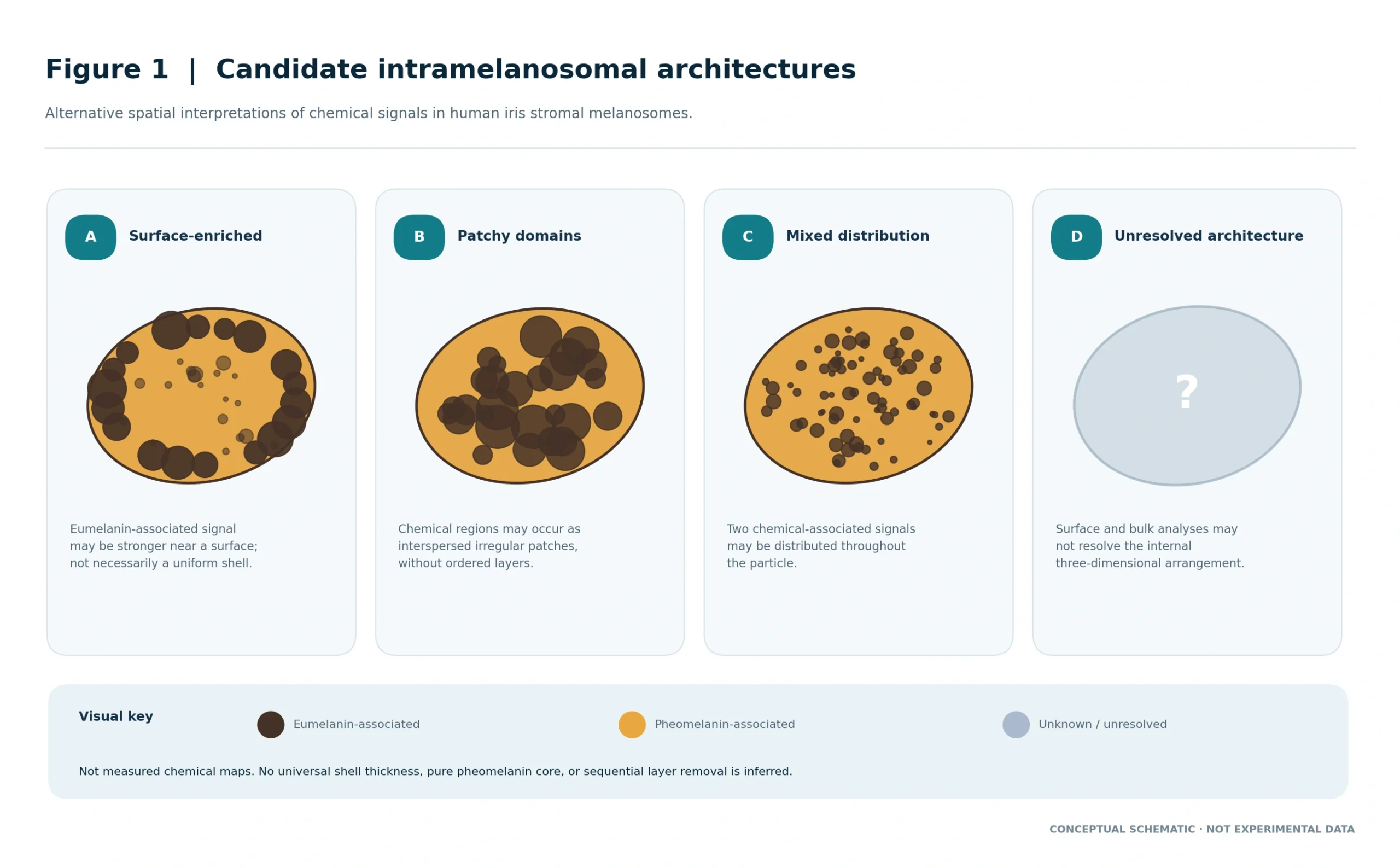
Peles et al. examined human iris stromal melanosomes from dark-brown and blue–green irides with bulk eumelanin:pheomelanin ratios of 14.8 and 1.3, respectively; surface-sensitive photoionization measurements were interpreted as revealing a common eumelanin-dominated outer signature [5]. This supports the possibility of chemically heterogeneous interiors with surface enrichment, but the study did not expose these granules to the proposed short-pulse challenge or establish that shell thickness controls any physical response. Surface-sensitive photoionization is not a three-dimensional chemical map. It does not determine a uniform shell thickness, a pure pheomelanin core, or an ordered layer-removal process. Nanoscale Cu/Ca/Zn mapping of purified mouse iridial granules revealed both peripherally enriched and more homogeneous distributions. Elemental markers are imperfect proxies for pigment chemistry, the tissue provenance is not identical to isolated human anterior stromal granules, and these observations do not constitute direct human eumelanin/pheomelanin mapping [6]. We will therefore compare surface enrichment, patchy organization, mixed distribution and indeterminate structure rather than assume one architecture.
Figure 1 illustrates alternative chemical-organization models compatible with differing surface- and bulk-sensitive observations without treating any schematic as a measured human iris stromal melanosome map.
2.3. Visible-wavelength response remains unverified
Peles and Simon investigated composition-related absorption in isolated iridal melanosomes in the ultraviolet [8,9]. Wavelength specificity prevents transfer of those coefficients or directional comparisons to 532 nm
without measurement. A 532-nm nonlinear-absorption analysis in a skin-laser experimental context and picosecond threshold studies using porcine eye-derived melanosomes demonstrate sensitivity to excitation regime and wavelength [10,11]. They do not isolate a human iris core–shell effect at nanosecond durations. Fluorescence-lifetime measurements on isolated bovine and porcine melanosomes also reveal evolving photophysical properties during extended 515-nm exposure over minutes [12]. They support the general idea that melanin state is not necessarily invariant, but their duration, species and illumination regime do not validate any sequential-exposure mechanism after LID.
Reviews of melanin photooxidation and analytical degradation markers provide methodological context for designing chemistry assays, not for assuming that a short clinical-wavelength pulse recreates a prolonged ultraviolet or visible-light exposure [13,14]. Additional primary evidence clarifies why a first-exposure explanation must be competitive rather than singular. RPE cell damage in a pulsed-laser experiment decreased in threshold with increasing pulse count [28], whereas single-microabsorber experiments demonstrated self-limited laser-induced vapor-bubble growth [31]. These are distinct observations—cumulative injury in cellular RPE versus transient bubble physics—not proof of progressive removal of a biochemical shell.
Moreover, photosensitized singlet-oxygen production at 532 nm in melanin solutions and melanocyte systems [29] cannot be equated with the response of isolated human stromal granules under a single laboratory challenge. More recent modeling and phantom studies underscore that absorber proximity and macroscopic fluence distribution can change predicted thermal and cavity responses even when pigment chemistry is not varied [32,33]. These are alternative-mechanism constraints, not measurements of human iris melanosomes.
2.4. Biochemical response is not the same as iris color response
A small rabbit iris laser experiment reported anterior stromal pigment reduction and increased melanophages at later histological assessment [15]. This motivates, but does not conclusively identify, separate early physical and later biological response phases. At the same time, stromal geometry, vascular responses, illumination, pigmentation depth and collagen scattering affect the visible phenotype. An organelle-level compositional effect may therefore exist without corresponding to a predictable human eye-color outcome, and the reverse is also possible: a visible change may occur without demonstrating any core–shell contribution [1,2,15,16].
2.5. Cross-scale phenotype is a constraint, not a chemical assay
At the tissue scale, the L-OAT phenotyping framework proposes recording optical–architectural differences, whereas LIPS provides a structured description of anterior stromal pigmentation [22,23]. These complementary descriptors may supply explanatory covariates for future clinical or explant investigations; neither resolves eumelanin/pheomelanin composition, melanosome surface enrichment or a shell thickness. An association between a visible grade and a physical-response endpoint would therefore be a cross-scale correlation, not proof of a chemical mechanism. The additional contribution proposed here is a donor-tested chemical variable measured independently from optical phenotype. Its predictive value should first be assessed against straightforward pigment-load, stromal- depth and reflectance models. Only a reproducible increment beyond these reference models would motivate an integrated organelle–tissue explanation.
Table 1. Evidence hierarchy and permissible inference
| Observation | Level of direct support | Not established |
|---|---|---|
| Eumelanin and pheomelanin differ among human iris preparations [3,4] | Human mixed iris material and cultured uveal cells | LID response thresholds or within-granule spatial maps |
| Common eumelanic surface in selected human granules [5] | Surface-sensitive measurements in selected human stromal granules | Universal shell, shell thickness or sequential removal |
| Heterogeneous elemental maps challenge simple casing [6] | Purified mouse iridial granules; indirect Cu/Ca/Zn proxy maps | Direct human chemical architecture or LID behavior |
| Melanin optics and state can vary [8–14] | UV organelle optics; skin/porcine/bovine laser models; chemistry studies and reviews | Human stromal 532-nm short-pulse core–shell effect |
| Delayed melanophage response after iris irradiation [15] | Rabbit iris histopathology | Composition-dependent human clearance or clinical benefit |
3. A discriminating, multi-route mechanistic hypothesis
The central hypothesis is that objectively measured organelle chemistry contributes information beyond conventional measurements of pigment quantity and anatomy. It does not require the existence of a complete physical shell. A surface-enriched eumelanin state, heterogeneous mixing, patchy domains or oxidation gradients are all experimentally distinguishable candidate architectures. The effect may be absent, limited to debris processing, or restricted to samples showing a particular surface state.
3.1. Route A: initial photophysical response
Under equal incident exposure, composition may affect response through absorption, nonradiative relaxation, local morphology or another mechanism. A first analysis will quantify the total association between independently measured chemistry and a physical endpoint. A second, deliberately narrower analysis will add a measured visible-wavelength absorption proxy; disappearance of the chemistry association after this adjustment is compatible with optical mediation, not evidence that chemistry is irrelevant. These are observational contrasts and cannot prove a particular molecular mechanism [8–11].
3.2. Route B: surface-state dependence after an initial perturbation
A first perturbation can alter the distribution and state of absorbers before a later challenge. The corresponding randomized history effect is broader than chemical change: it may reflect loss of the most reactive particles, fragmentation, aggregation, heating, residual bubbles or optical realignment. Surface chemistry becomes a specific candidate only if a reproducible state difference is measured independently and simpler causes are examined. No direction of the second response is assumed.
3.2.1. The first exposure can change the target without proving shell removal
A history-dependent second response can arise whenever the first encounter changes the distribution of absorbers, the surrounding medium, or the instrument-observed endpoint. In retinal models, vapor microbubbles around laser-heated melanosomes are directly observed, and RPE injury thresholds have been shown to depend on pulse number [27,28]. These results demonstrate that repeat-exposure behavior requires physical explanation; they do not identify surface chemistry as the cause in human iris melanosomes. An attenuation of subsequent signal may simply reflect destruction or removal of the most absorbing particles; an increase may reflect aggregation, a changed optical path, or a cumulative injury threshold. The term “first exposure” should therefore designate a defined ex vivo perturbation and observation interval, not a presumed biochemical peeling event. The proposed core–shell hypothesis does not predict a necessary direction of change. In particular, a common eumelanic surface [5] does not establish that one pulse removes it, exposes pheomelanin, reduces absorption, or makes later pulses more effective. Each transition would require direct post-exposure structural and chemical measurement.
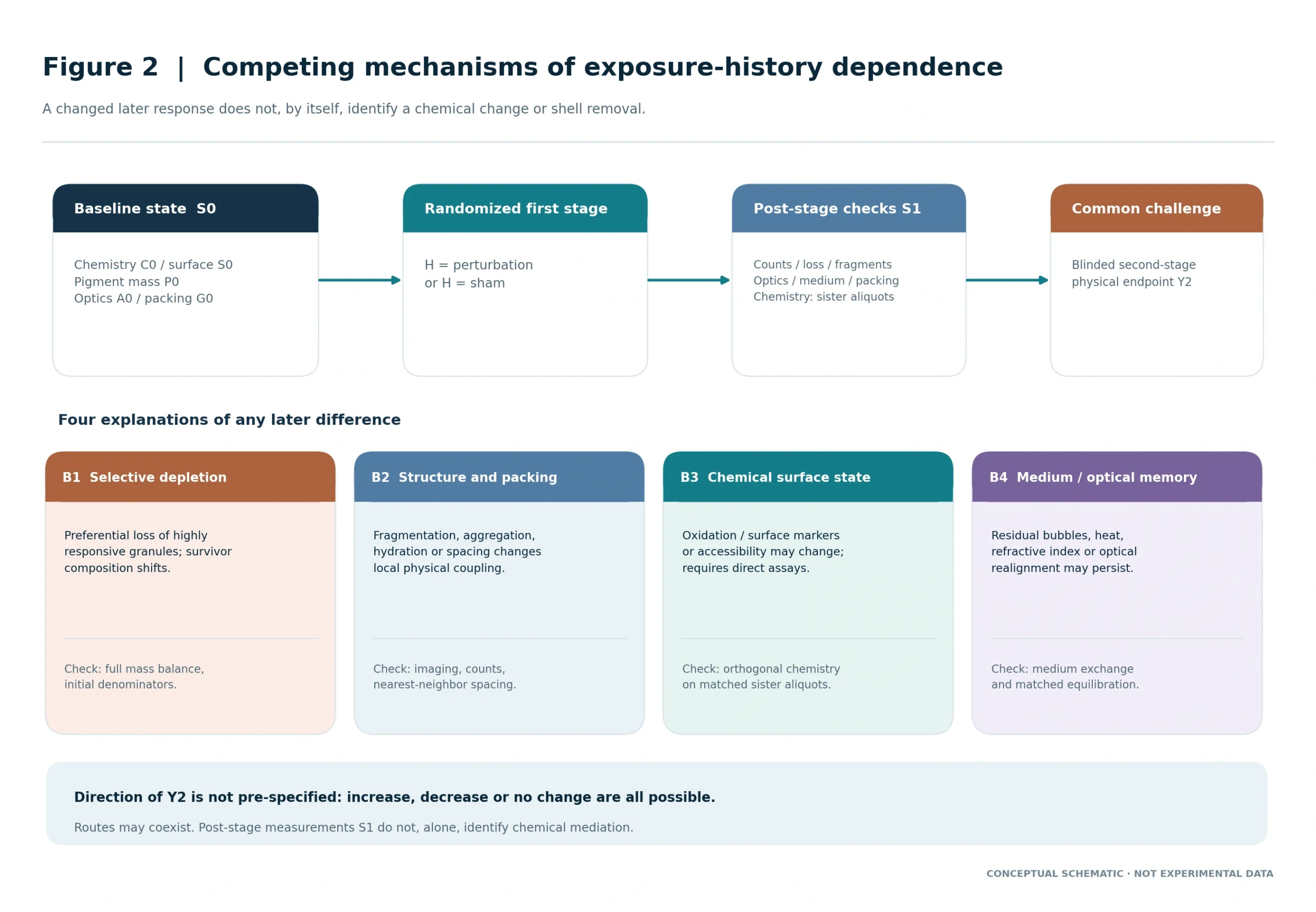
3.2.2. Four competing routes to a changed second response
Route B1—selective depletion and survivor enrichment: the first perturbation disproportionately changes or eliminates the most responsive granules, leaving a population with a different average absorption, size, and geometry. The second challenge then appears weaker even though the chemistry of each surviving organelle has not changed. Whole-sample mass balance and particle survival counts should be reported alongside within- survivor measurements.
Route B2—structural or colloidal rearrangement: fragmentation, local cracking, membrane damage, decompaction, hydration shifts, or reversible aggregation may change the effective interaction cross-section or microscopic scattering. These mechanisms can alter signals without a new eumelanin:pheomelanin ratio. Because short-pulse bubble nucleation is strongly affected by particle heating and its aqueous microenvironment [27,31], a photoacoustic amplitude change cannot be interpreted as surface-chemistry change in isolation. Route B3—chemical surface modification: a measurable change in oxidation markers, metal association, surface ionization signature, or access to previously buried pigment domains could change optical and redox properties.
Melanin photooxidation and visible-light photosensitization have been demonstrated in non-iris systems [13,14,29], and pump–probe signatures can change irreversibly following intense irradiation of iron- associated model eumelanin [30]. Yet prolonged solution illumination, near-infrared pump–probe photodamage, and clinical-wavelength nanosecond iris exposure are not equivalent regimes. This route is a specific proposed mechanism, not an established effect of first-pulse LID. Route B4—medium and transient optical memory: residual gas bubbles, local heating, changing refractive index, suspension motion, or evolving illumination geometry may briefly modify the next measurement without any persistent change to the melanosome. Controlled medium exchange, thermal equilibration and time-separated repeat observations are required before labeling the difference an intrinsic particle memory.
Self-limited microbubble dynamics around model microabsorbers demonstrate why the fluid-phase response deserves its own endpoint [31].
Figure 2 separates the randomized first-stage history effect from four competing, potentially coexisting explanations of the later physical response.
3.2.3. Timing and environmental memory
Heat deposition, microbubble dynamics and fragmentation may occur during or shortly after a single event, whereas chemical alterations can follow different kinetics and macrophage handling requires a cellular context. These processes must not be arranged as a single mandatory biological sequence. We propose time-separated readouts that verify return of medium and optical baselines before comparing persistent particulate states; recovery of a physical signal would not imply restoration of an eumelanic shell [12,27,28,31].
3.2.4. An explicit state-transition prediction
Let S0 denote prespecified baseline attributes: chemical profile, surface signal, particle number and size, packing, visible-wavelength optics, and medium conditions. Let S1 denote the measured post-first-stage state including recovered and lost material; H denotes randomized first-perturbation versus sham assignment; Y2 denotes the later blinded physical endpoint. The randomized contrast H → Y2 estimates a total history effect. The association between S1 and Y2 is a mechanistic clue, not an identified mediation effect, because S1 is affected by H and may be measured on selected survivors. Spatial shell removal requires a separate localization experiment.
3.3. Route C: fragmentation and phagocytic processing
If two characterized organelle populations yield fragments with different redox signatures or surface chemistry at the same physical size and loading, downstream pigment uptake and processing could differ. The outcome must be measured directly in appropriate cell models; observed melanophages alone cannot establish a difference caused by pigment composition. Particle exposure is also potentially detrimental to ocular outflow biology, so no favorable physiological consequence can be presumed from greater clearance [15,19]. P- CLEAR is relevant here only as a conceptual reminder to measure released material, residence/processing and recovery as distinct phenomena; it supplies no evidence for a composition-dependent clinical clearance advantage [24].
3.4. A formal statement without unsupported kinetic constants
The model uses baseline chemistry C0, surface marker S0, optical measure A0, pigment mass P0, geometry/packing G0, randomized history H and the later response Y2, with donor and preparation batch explicitly clustered. The primary prediction is incremental out-of-donor information from measured chemistry beyond P0, G0 and A0. The randomized H contrast and C0 × H effect modification answer separate questions.
No effect size, direction, rate constant or clinical dose is assumed. Iris color is a tissue phenotype and cannot substitute for Y2.
3.5. Joint chemistry–packing model: when two explanations may interact
The distinction is between three candidate pathways: (i) C0 predicts Y2 through optical absorption A0; (ii) packing G0 modifies local heating and response even when chemistry is constant; and (iii) H changes the distribution or intrinsic state of absorbers, possibly in a chemistry-dependent manner. We will report baseline predictors and post-first-stage measurements separately. A chemistry × packing interaction is an exploratory effect-modification test, not a presumption that one pathway dominates [10,11,32,33]. A 2026 three-dimensional thermal-modeling study of epidermal melanosome clusters predicted spacing- dependent thermal cross-talk and cooling kinetics in simulated clusters after 532-nm, 10-ns heating; it did not directly measure human iris organelles [32].
A separate 2025 melanosome-containing optical phantom study linked the distribution of cavitation to local fluence and spot geometry [33]. Neither experiment tested human iris stromal chemistry or validated a clinical procedure. Their significance here is to require measurement of nearest-neighbor distance and cluster architecture rather than assuming that every post-exposure change represents new pigment chemistry. Accordingly, isolated-granule studies should quantify or constrain local spacing, and tissue explants should test whether any signal remains when native stromal geometry is restored. An association that vanishes when packing is measured supports a simpler structural explanation. A reproducible chemical association within comparable packing strata deserves additional testing, but remains distinct from a claim of shell disruption.
3.6. Appearance and causal inference are different measurement problems
An independent warning comes from human skin: Omatsu et al. reported ultrastructural melanosome disruption in human skin even in some sites without immediate visible whitening after picosecond exposure [34]. This is evidence of measurement dissociation in a different tissue and pulse regime, not a surrogate for human iris damage or surface chemistry. The proposed experiment records visible signal, microscopic disruption, recovered pigment mass and chemistry separately.
3.7. Optical mediation and randomized history effects
A reproducible chemistry–response association may be mediated by visible-wavelength absorption. Adjusting for A0 asks whether any association remains beyond that optical pathway; it does not estimate the total chemical contribution. Conversely, persistence after adjustment does not identify a direct surface mechanism because unmeasured structure can affect both chemistry markers and the endpoint. We will report total prediction and optical-conditioned prediction side by side, with clearly different interpretations. Randomization of first-stage assignment H identifies the total effect of that experimental history at the preparation level under adequate allocation and follow-up. It does not randomize chemical state or eliminate depletion. A baseline C0 × H interaction tests whether the effect differs by pre-exposure chemistry.
Post-state surface assays on randomized sister aliquots may support a material change, but cannot show that the same individual granule chemically converted unless a valid within-particle method is available. Decision logic is prespecified: (a) chemistry predicts response, but only through A0—an optical explanation remains; (b) H changes Y2 without chemical-state evidence—a general physical history effect remains; (c) C0 × H replicates and orthogonal state assays change—a chemistry-linked mechanism warrants follow-up but is not proven; (d) chemical domains are directly localized before and after exposure in appropriate material— only then may a spatial surface-change claim be considered. In every case particle loss, packing and medium effects must be accounted for [5,6,12,27,31–33].
4. Falsifiable predictions
Table 2. Pre-specified predictions and observations that would weaken them
| Prediction | Supportive observation | Disconfirming or limiting observation |
|---|---|---|
| P1 · Composition adds information | Donor-held-out predictive improvement beyond load, geometry and optical baseline | No reproducible incremental prediction after matching and adjustment |
| P2 · Exposure-history contrast and chemical effect modification | Randomized first perturbation changes later response; evaluate chemistry-by-history interaction and measured post-state chemistry separately | Explained by depletion, medium or geometry; no replicated chemical interaction |
| P3 · Chemistry alters debris handling | At matched size and load, characterized debris differs in uptake or processing | Fragment burden and morphology account for the difference |
| P4 · Optics and appearance can dissociate | Explant reflectance and tissue scattering do not follow pigment mass or disruption one-to-one | Simple pigment-mass-and-depth model explains changes |
| P5 · Human translation is not automatic | Independent donor and explant analyses reproduce organelle association | Effect vanishes in intact stroma or is non-human only |
| P6 · Chemistry × packing is distinguishable | Replicated chemistry-by-packing term improves donor-held-out predictions or packing-stratified results | Packing, dose or survivor enrichment explains the result |
5. An executable research program
5.1. Phase 0 — Establish assay feasibility and contamination controls
Secure ethically sourced human donor iris tissue with documentation of donor provenance, handling time, ocular history where available, storage conditions and processing batch. Use tissue-layer dissection and orthogonal imaging to distinguish anterior stromal melanosomes from posterior iris pigment epithelium, cellular debris and other pigment-bearing compartments. Prior to hypothesis testing, quantify assay repeatability and the degree to which extraction alters particle surfaces, aggregation or chemical yield. If these controls fail, an inferred core–shell effect would be uninterpretable and the program should not advance to laser-response comparisons.
5.2. Phase I — Independent biochemical and structural phenotyping
Estimate eumelanin- and pheomelanin-associated marker profiles using validated chemical-degradation and chromatographic methods (for example, PTCA/PDCA for selected eumelanin-associated products and TTCA/4-AHP-related markers for selected pheomelanin components), with assay-specific interpretation, blanks, recovery controls and reference standards [13,14]. Ratios are assay-dependent proxies rather than a direct map of an intact organelle. Combine these bulk measurements with complementary microscopy and surface-sensitive assays; where material permits, test spatial localization with an orthogonal depth-resolved or correlative method. Classify architecture as measured surface-enriched, ambiguous or incompatible with a simple casing interpretation. Avoid assigning shell thickness without direct spatial evidence. A key design feature is sampling across donors and regions, not only across visually classified eye colors. Document organelle dimensions, number density, aggregates, regional stromal depth and whether measurements are performed on individual particles or pooled extracts. The donor, not each granule, must remain the primary biological sampling unit for population-level inference.
5.3. Phase II — Wavelength-specific optical characterization
Measure wavelength-resolved optical properties in the visible region, including the experimental wavelength of interest, in a configuration that separately evaluates absorption-related and scattering-related signals where feasible. Calibrate instrument response and document how suspension concentration, particle aggregation, solvent and optical path length affect estimates. Ultraviolet results [8,9] must remain mechanistic context and cannot be substituted for these observations. For exploratory energy-deposition comparisons, explicitly report whether models use delivered incident exposure or a measured/estimated absorbed-dose proxy, because conditioning on absorption can remove a genuine optical mediating pathway.
5.4. Phase III — Controlled ex vivo response and state-transition tests
A feasibility pilot will select and lock one objectively reproducible primary physical outcome before confirmatory samples are analyzed. The preferred candidate is second-challenge incremental microscopic disruption in prespecified fields, assessed by blinded imaging and reconciled to particle counts and recovered fragment mass; if this cannot be measured reliably, an independently validated optical/acoustic endpoint must be specified instead. The endpoint is quantified per preparation and analyzed across independent donor eyes— not as hundreds of independent granules. Study exposures require instrument calibration, but no clinical energy, spot or treatment protocol is inferred [10,11,20]. Each donor supplies comparable aliquots assigned to first-perturbation or sham handling, followed by a separately characterized challenge; a subset also tests medium replacement and time-matched retention. Record first-stage loss, fragmentation, thermal state and particle distribution before challenge 2. Any dose or timing used in the ex vivo apparatus is documented for reproducibility but is not an implied patient-facing setting. The phase separation is consistent with TRA, which organizes response timings without validating a molecular mechanism [25].
5.4.1. Within-donor challenge arms and negative controls
Randomize comparable within-donor preparations to first perturbation or sham; include preassigned observation regions, a chemistry-only aliquot, and a probe-only negative control. A subsequent independent challenge is applied under the same calibrated readout configuration. Preserve the original preparation denominator and report first-stage attrition even when no intact target remains for challenge 2. Blinded assessment and independent donors, rather than multiple images of one donor, determine replicability. The primary randomized history contrast is calculated at the assigned preparation level without restricting analysis to visible survivors or conditioning on post-first-stage depletion. Secondary material-normalized and survivor-restricted analyses are useful mechanistic descriptions but can introduce selection or mediation bias. Present them as such, alongside whole-sample mass balance.
5.4.2. Four deliberately separable measurement layers
Layer 1—material balance: retained pigment mass, total intact particle count, fragment number and size distribution, plus discarded material. Layer 2—optical properties: wavelength-specific absorption-related proxies, reflectance/scattering and any observed shielding or aggregation. Layer 3—chemical state: independently validated surface-sensitive signatures, oxidation-associated markers and metal-binding covariates, without presuming a universal shell thickness. Layer 4—physical challenge outcome: pre-specified, reproducible signal or event classification, including microscopic displacement, photoacoustic response or cavitation when measurable. None is a surrogate for another. For a proposed physical endpoint, diagnostic readout stability must be validated independently from the perturbation under study. For example, a falling photoacoustic amplitude could reflect lower absorbed energy, a changed detector geometry or fewer absorbers rather than a decline in microcavitation. Conversely, a visible bubble or strong acoustic signal is not a direct measure of biochemical depletion. Microscopy and orthogonal optical diagnostics should confirm endpoint concordance.
5.4.3. Persistent state versus transient medium effects
Repeat the later challenge after predefined equilibration conditions using separate matched aliquots, including a controlled-medium-exchange arm and time-matched sham handling. Measure medium temperature, baseline optical response and particle position before challenge. A difference that vanishes after equilibration favors transient medium or thermal explanations; a persisting difference that correlates with independent chemical changes supports further testing of an intrinsic state mechanism, but remains insufficient to establish it alone. Temporal comparisons should avoid treating the same granule as intact after a destructive event.
5.4.4. Causal discrimination without assuming a photobleaching mechanism
Predicted signatures are distinguishable but need not be mutually exclusive. Depletion predicts loss of highly reactive absorbers; geometry predicts changes linked to fragmentation or packing; optical mediation predicts correspondence with independently measured absorption; transient-medium effects predict attenuation after verified equilibration; and chemical change requires repeatable, orthogonal state assays. If the data support several routes, the causal contribution of each may remain nonidentifiable. The result should be reported as mixed rather than forced into a single explanation.
5.4.5. Bias from repeated measurement of survivors
An analysis restricted to melanosomes still visible after the first perturbation is vulnerable to survivor selection: those lost or disrupted may have had systematically different baseline chemistry or optical responses. Pre-assign intention-to-observe sample regions; report the denominator of particles before each exposure, disappearance/fragmentation as separate events, and sensitivity analyses treating attrition as an outcome. The principal independent unit remains the tissue donor, not the number of granules or frames.
5.4.6. Manipulate packing as a competing cause rather than a nuisance variable
Use comparable donor preparations in which granule concentration, clustering state and aqueous conditions are quantified independently from chemistry. Where preparation methods allow, evaluate dispersed and characterized clustered aliquots while monitoring whether the manipulation itself changes aggregation, hydration, surface markers or measured absorption. This is a conceptual experimental contrast, not a claim that intact iris spatial architecture can be reproduced by a suspension. Reserve the clinically relevant optical geometry for later intact-stroma validation. Report granule size, concentration, nearest-neighbor spacing and fraction of aggregates, with native stromal depth added in explants. If manipulating packing also changes particle surfaces or optical absorption, the manipulation is not a clean isolation of geometry and must be interpreted cautiously. The 2026 modeling work illustrates a plausible nonchemical effect of spacing on thermal relaxation; it is not an empirical iris validation [32].
5.4.7. Distinguish organelle tracking from destructive chemistry assays
Within-organelle repeated measurement is attractive but cannot be assumed feasible for all assays. Many established chemical-degradation procedures consume the material; destructive surface assays may sample a different organelle population from the one generating a later physical response. The design should therefore combine nondestructive longitudinal readouts on immobilized or otherwise identifiable granules, if feasible, with randomized sister aliquots for destructive chemistry. These two streams answer related but nonidentical questions and should not be described as direct before-and-after chemistry on the same particle. Document identifiability, dropout and measurement-induced perturbation. For an ostensibly diagnostic optical probe, include probe-only controls, repeatability limits, blinded endpoint classification and time-matched sham handling. Count disintegrated or lost particles explicitly rather than restricting the analysis to surviving granules. A changing average spectrum in the remaining population can be caused by selection even when no individual granule has chemically transformed.
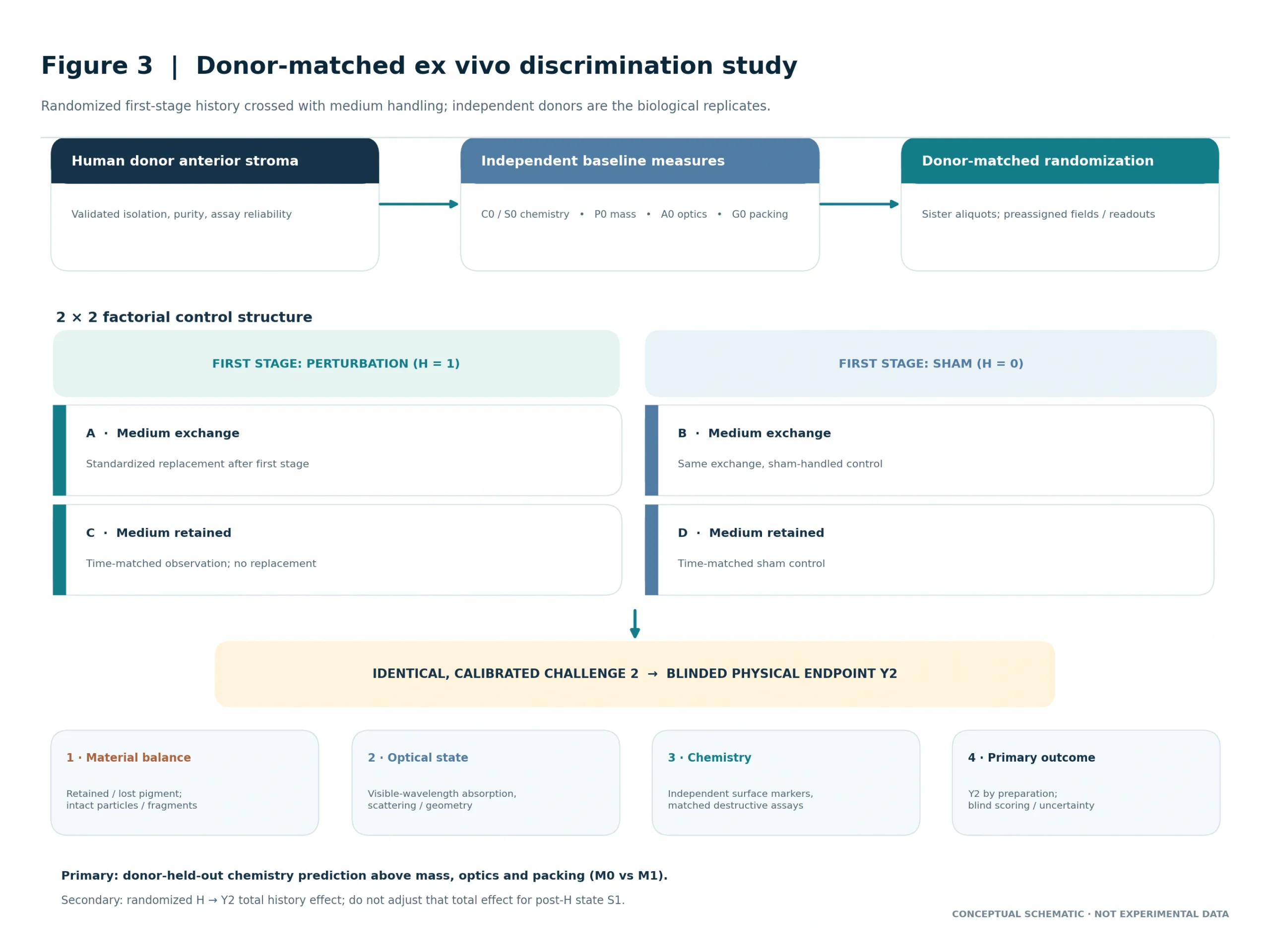
5.4.8. A factorial medium-exchange and observation-interval control
Use a prespecified 2 × 2 control structure: first perturbation versus sham, crossed with standardized medium exchange versus time-matched retention. Add a probe-only negative control and an optical realignment check.
Record temperature, visible or optically detected bubble persistence, particle concentration and motion before challenge 2. These measurements test environmental carryover without presuming a fixed relaxation interval [27,31]. If a response difference disappears after exchange or realignment, transient optical/medium memory is favored. If it persists but particle composition changes mainly through loss of high-response granules, survivor selection remains a competing explanation. If it persists with stable recovery accounting and concordant chemical-state measurements, the chemical hypothesis becomes more plausible—not proved. The exchange experiment can alter particle handling and thus requires its own sham and recovery controls.
Figure 3 summarizes the proposed donor-matched 2 × 2 perturbation-by-medium-handling design, the standardized second challenge and the independent measurement streams.
5.5. Phase IV — Iris explants and downstream particle handling
Any effect detected in isolated material should be challenged in human stromal explants, where depth, collagen architecture, local scattering and melanosome packing can dominate delivered energy [1,2,16]. Separately quantify standardized reflectance, pigment mass, viable tissue injury, fragment characteristics and, where available, biological uptake in an independently validated macrophage-related model. No one measure should be treated as a surrogate for all others. The presence of pigment-laden macrophages should not be interpreted as proof that compositional variation improves clearance or ocular safety [15,19].
5.6. Phase V — An exploratory clinical-phenotype bridge
Clinical translation should be considered only after replicated organelle and explant results. A human polarization-sensitive anterior-segment OCT study explored an imaging-derived iris melanin concentration estimate and its association with colorimetry in brown irides [17]. This is an indirect tissue-scale imaging proxy rather than a chemically validated measurement of absolute melanin mass. This technique does not distinguish eumelanin from pheomelanin or resolve a melanosome core–shell phenotype. A future retrospective imaging analysis could test whether baseline stromal imaging or colorimetric patterns predict longitudinal change, with device settings, region, pupil size, follow-up, treatment exposure and image white balance recorded. Such correlations would not validate the underlying chemistry without tissue-based calibration. LIPS and L-OAT variables could be prospectively recorded as tissue-scale descriptors, but must not be used as substitutes for chemical melanosome measurements [22,23].
Table 3. Proposed study sequence and primary interpretation rule
| Phase | Output | Gate before proceeding |
|---|---|---|
| 0 · Material validity | Purity, processing artifacts, assay repeatability | Do not infer surface chemistry if extraction changes it |
| I · Chemistry and architecture | Bulk ratio and independent surface phenotype | No shell claim without spatial evidence |
| II · Optical baseline | Visible-wavelength absorption and scattering proxies | Separate incident from absorbed exposure |
| III · Physical perturbation | One blinded endpoint, material accounting, randomized sham-first contrast | Measurement validated; independent donor replication |
| IV · Tissue and cell context | Explant transfer, fragment processing and injury | Separate physical response from safety and clearance |
| V · Clinical imaging, optional | Prospective or retrospective association only | No inference of melanosome chemistry from iris color |
6. Statistical analysis and adjudication of mechanisms
Two analyses should be prespecified, with one confirmatory primary claim. The preferred primary analysis tests whether C0 or a declared surface marker adds donor-held-out predictive information for the blinded physical outcome beyond baseline pigment mass, geometry and wavelength-specific optical measurements (M0 versus M1). The randomized H → Y2 contrast is a key secondary analysis; C0 × H and post-state mechanistic associations are exploratory until a feasibility pilot supports adequate independent donor numbers.
Predictions must be evaluated at donor level, never by random splitting of images or particles from the same donor. Specify a primary chemistry feature, one physical endpoint, a clinically neutral material/sample unit and the minimum detectable scientifically relevant difference before confirmatory work. Use donor-level clustering in mixed models, and batch-level terms when appropriate. Report cross-validated error or calibration where applicable, effect estimates with uncertainty intervals, and missing/failed assay rates. Pilot-derived between- donor variation and measurement repeatability—not particle count—should determine sample size. A non- significant result with a wide interval is inconclusive, not evidence that chemistry has no effect. Distinguish baseline prediction, randomized total history effect and post-exposure mediation: these analyses do not share a causal interpretation. If composition loses incremental value after absorption is included, the result is compatible with optical mediation. If a chemical surface marker remains associated, confounding by unmeasured morphology is possible. Measuring S1 after H cannot, without additional assumptions or interventions, quantify a causal chemical mediation proportion.
6.1. Separate total history dependence from chemical mediation
Estimate the total randomized H → Y2 contrast using all assigned preparations, including samples with first- stage particle loss. Do not adjust the primary total contrast for P1, S1, A1 or post-H packing, because these may be consequences of H. Their introduction belongs to explicitly exploratory mechanism models, in which mediator–outcome confounding and selection must be acknowledged. Test one prespecified C0 × H interaction only if pilot data justify donor-level precision. The interaction reflects effect modification by measured baseline composition, not conversion of chemistry. Independent paired aliquots may demonstrate changes in chemical-state distributions, but attributing these changes to within- particle conversion requires independent spatial or time-resolved evidence and accounting for lost material.
6.2. Disconfirming signatures and reporting rules
The chemistry-specific pathway is weakened if surface markers are not reproducible, extraction changes their direction, the putative effect disappears with appropriate mass/geometry controls, donor-held-out prediction fails, or the signal is absent in intact iris explants. Medium equilibration or sham processing that reproduces the signal instead favors an environmental or assay explanation. A negative claim requires adequate donor- level precision; otherwise the finding should be described as indeterminate.
6.3. Pre-specified interaction tests and levels of inference
The analysis has three separate estimands. A: incremental donor-held-out prediction by independently assayed C0/S0 over baseline pigment mass, optical measures and packing. B: the preparation-level total effect of randomized H on Y2, without post-exposure adjustment. C: exploratory C0 × H effect modification under baseline covariate control. Estimand C does not test whether an individual melanosome acquired new chemistry; that requires an independent post-state assay stream. For continuous Y2, use a donor- and batch-aware model with stated diagnostics; for a binary event, a hierarchical generalized model is appropriate. Constrain the confirmatory feature set to one primary chemical variable and predefined physical endpoint; treat additional spectroscopy bands and packing interactions as exploratory. Report donor-held-out predictive intervals and appropriate uncertainty rather than a granule-level p-value. The sample size must reflect independent donors rather than observations per donor. Describe post-state distributions (S1, retained mass, aggregation and optics) as potential intermediates. Survivor-restricted analyses and statistical adjustments for post-H variables can bias estimates of the total randomized effect. If direct mediation is claimed in later research, an intervention on the candidate mediator or a defensible identification design is needed. Replication in intact human stroma remains a separate translational test.
6.4. Decision gates: supportive, restricted, or null outcomes
A supportive outcome for baseline chemistry requires a reproducible gain in donor-held-out prediction above mass, measured optics and packing, with reliable chemistry assays. A supportive history effect requires a randomized, preparation-level Y2 contrast with complete loss/fragment accounting. A chemistry-specific history interpretation additionally requires a replicated C0 × H result plus independent chemical-state evidence and plausible exclusion of nonchemical causes. If only optics or packing predicts response, report the restricted model. If uncertainty cannot exclude meaningful effects, report an inconclusive rather than null result. Importantly, no fixed numerical cutoff, effective clinical dose or expected color outcome is inferred from studies of isolated particles, skin models, retinal pigment epithelium or optical phantoms. Assay reproducibility, a feasible donor sample size and an ethical donor-tissue protocol are prerequisites to any quantitative hypothesis test. Any future in vivo validation would require its own clinical, safety and governance design.
6.5. A graded evidentiary ladder rather than a binary positive result
An ordered evidentiary ladder prevents overinterpretation: Level I, reproducible incremental prediction by baseline chemistry; Level II, a randomized preparation-level history effect; Level III, donor-replicated baseline chemistry × history modification; Level IV, independent biochemical or surface assays documenting altered material state with adequate recovery controls; Level V, localized spatial evidence linking a specific altered surface domain to the response. Higher levels are not implied by lower levels, and Level IV need not establish Level V. A clinical advantage is not an outcome at any level. Before pilot reliability is known, the confirmatory target is Level I; Level II is a prespecified secondary randomized contrast. Levels III–V define mechanistic follow-up, not presumed findings. Failure of Level I under precise conditions narrows the value of the chemistry-prediction hypothesis. Failure of Level IV weakens the remodeling branch but does not rule out purely optical chemistry-related effects.
Figure 4 distinguishes baseline prediction, randomized history effects, effect modification, independently measured material-state changes and spatial localization as separate evidentiary levels.

6.6. Pre-registration, measurement reliability and transparent stopping rules
Preregister one physical endpoint, one primary chemistry feature, the donor-level analysis unit, M0/M1 covariates, one primary model-comparison metric, missing-data rules and a scientific minimum-effect criterion. A pilot should estimate assay repeatability and independent-donor variance; no sample-size figure is invented here. Lock criteria before confirmatory data collection, and archive outcome definitions, failed assays and analysis-code versions. Clinical efficacy and procedural safety are not tested. Pause or narrow inference if the isolation process alters surface signatures, the readout itself changes particles, chemistry assays are not reproducible, the planned donor sample cannot provide adequate precision, or the whole-sample material balance is unreliable. A reproducible optical or packing-only explanation is a valid scientific result. Null and mixed results should be reported with full tested contrasts.
7. Major alternative explanations and likely failure modes
The main competitors are pigment quantity and stromal depth; local packing, absorber spacing and optical shielding; first-stage particle depletion and survivor selection; thermal or fluid-phase memory; and extraction- induced changes in surface state. Tissue color, delayed melanophage activity and donor-dependent phenotype introduce separate cross-scale uncertainties [1,2,15,16]; non-iris optical and packing experiments provide additional but indirect constraints [12,20,32,33]. Each competitor has a corresponding prespecified control: mass balance, calibrated absorption, imaging of particle neighborhoods, medium exchange, probe-only testing, or tissue-explant replication. An additional failure mode is equating initial visible whitening or a change in optical signal with chemical photobleaching. Short-pulse skin observations show that vacuoles and immediate visible whitening may dissociate from microscopic disruption [34]; visible-light photosensitization and pump–probe photodamage studies involve distinct cellular or model-melanin settings [29,30]. Either observation can occur without establishing surface-eumelanin loss in an iris melanosome. Human iris work must therefore keep optical
whitening, mass depletion, surface chemistry, and later tissue appearance as independent observations. Omatsu et al. directly found that melanosome disruption in human skin may occur without obvious immediate whitening [34]; conversely, that finding provides no permission to infer microscopic iris disruption from visible surface changes. Physical endpoints must be independently verified. Importantly, the same observations can have different biological meanings. Greater physical fragmentation is not necessarily a favorable endpoint, because liberated particles can also contribute to obstruction, inflammation or secondary pressure elevation. A published severe pigmentary-glaucoma case after cosmetic laser iris treatment demonstrates why physical disruption, pigment clearance, ocular safety and functional outcome must be treated as separate domains; a case report cannot estimate incidence [19]. This hypothesis cannot justify changing clinical laser settings or claiming greater efficacy in any iris-color category. L-SAFE provides a pre-exposure assessment framework for considering safety states, not a method of validating the present physical-response hypothesis or an endorsement of intervention [26].
7.1. Specific objections a critical reader should be able to test
A critical reader may object that (i) a common surface signature is not a uniform shell; (ii) optical absorption or packing alone explains response; (iii) later responses reflect selection or microbubble persistence; (iv) destructive assays cannot follow the same particle; and (v) isolated granules do not reproduce intact iris stroma. We address these objections respectively by orthogonal spatial measurement, calibrated optical and geometric baselines, full material accounting and medium controls, explicit separation of tracking versus destructive sister-aliquot chemistry, and human explant validation [5,6,10–12,27,31–33]. A stronger physical disruption signal also cannot be translated into better cosmetic results, more efficient pigment clearance or ocular safety. The current article proposes no clinical parameter change. Secondary pigmentary glaucoma after cosmetic iris laser procedures has been reported; this possibility strengthens the need to keep physical response, downstream processing and clinical safety separate [19].
7.2. Evidence concerning chemical photodamage must not be over-translated
Oxidatively modified synthetic Dopa-melanin differs from a native iris stromal melanosome exposed to a nanosecond visible-wavelength pulse; it illustrates how chemical-state measurements might be designed, not what SSMM necessarily does [35]. Likewise, time-varying fluorescence after prolonged 515-nm exposure in bovine/porcine material cannot be used to claim rapid iris surface remodeling [12].
8. Position relative to other proposed response frameworks
SSMM describes the intended iris-stromal anatomical context and response-guided conceptual approach; the present study does not claim that any specific melanosome chemistry is established by SSMM [21]. The distinction is between the tissue selected for investigation and the chemical organization of the organelles within that tissue. This paper concerns only the latter question. At the baseline tissue-phenotype level, L-OAT frames spatial and optical architecture, and LIPS structures clinical descriptions of anterior stromal pigmentation [22,23]. Neither is a chemical classification. A falsifiable bridge would ask whether independently measured human-donor biochemical/surface features improve the explanation of ex vivo optical or physical response after accounting for those tissue-scale descriptors; matching clinical color classes alone would be inadequate. At the post-exposure level, P-CLEAR separates released pigment and subsequent compartmental processing, while TRA separates response domains and time courses [24,25]. These frameworks can inform the selection and timing of outcomes, but do not demonstrate that a core–shell architecture exists, controls disruption, or improves clearance. A time-dependent observation without a measured chemistry × state interaction would be compatible with simpler depletion and recovery explanations. L-SAFE concerns pre-exposure safety assessment [26]. It is relevant only as a boundary: any organelle-scale physical difference must remain distinct from inflammation, trabecular effects, corneal/endothelial status and
clinically meaningful safety. No result in this hypothesis could replace standard safety assessment, and a greater physical response is not synonymous with a better outcome. These frameworks organize different levels of observation; none independently validates the proposed molecular mechanism. The present study is falsifiable even if subsequent data support only pigment load, optical absorption or packing as explanations.
9. Conclusions
Human tissue and cell studies establish chemical heterogeneity of iridal melanin, while a selected human stromal melanosome study reports a eumelanin-associated outer signature [3–5]. This does not demonstrate a universal core–shell geometry or a 532-nm nanosecond chemistry effect. Evidence from other species, tissues and pulse regimes motivates controlled experiments but cannot substitute for human-donor organelle and explant tests [6,8–14,27,32–35]. The proposed contribution is a donor-replicated test of baseline chemistry as an incremental predictor, combined with a distinct randomized exposure-history contrast. A later-response change, even if substantial, does not itself establish chemical remodeling. That stronger claim requires reliable material accounting, orthogonal chemical assays and exclusion of depletion, geometry and transient optical effects; a claim of spatial surface transformation requires direct localization. Restricted optical, packing-only, mixed and precise null results would remain scientifically informative. This work specifies an experimental hypothesis, not a change in clinical laser practice, nor evidence of cosmetic efficacy or ocular safety. Associated Lumineyes frameworks provide relevant anatomical, phenotyping, temporal and safety context without validating the mechanism [21–26].
References
- Wilkerson CL, Syed NA, Fisher MR, Robinson NL, Wallow IH, Albert DM. Melanocytes and iris color: light microscopic findings. Arch Ophthalmol. 1996;114:437–442. doi:10.1001/archopht.1996.01100130433014. PMID:8602782.
- Imesch PD, Wallow IHL, Albert DM. The color of the human eye: a review of morphologic correlates and of some conditions that affect iridial pigmentation. Surv Ophthalmol. 1997;41(Suppl 2):S117–S123. doi:10.1016/S0039-6257(97)80018-5. PMID:9154287.
- Prota G, Hu DN, Vincensi MR, McCormick SA, Napolitano A. Characterization of melanins in human irides and cultured uveal melanocytes from eyes of different colors. Exp Eye Res. 1998;67:293–299. doi:10.1006/exer.1998.0518. PMID:9778410.
- Wakamatsu K, Hu DN, McCormick SA, Ito S. Characterization of melanin in human iridal and choroidal melanocytes from eyes with various colored irides. Pigment Cell Melanoma Res. 2008;21:97–105. doi:10.1111/j.1755-148X.2007.00415.x. PMID:18353148.
- Peles DN, Hong L, Hu DN, Ito S, Nemanich RJ, Simon JD. Human iridal stroma melanosomes of varying pheomelanin contents possess a common eumelanic outer surface. J Phys Chem B. 2009;113:11346–11351. doi:10.1021/jp904138n. PMID:19618947.
- Gorniak T, Haraszti T, Suhonen H, et al. Support and challenges to the melanosomal casing model based on nanoscale distribution of metals within iris melanosomes detected by X-ray fluorescence analysis. Pigment Cell Melanoma Res. 2014;27:831–834. doi:10.1111/pcmr.12278. PMID:24903463.
- Anderson RR, Parrish JA. Selective photothermolysis: precise microsurgery by selective absorption of pulsed radiation. Science. 1983;220:524–527. doi:10.1126/science.6836297. PMID:6836297.
- Peles DN, Simon JD. The ultraviolet absorption coefficient of melanosomes decreases with increasing pheomelanin content. J Phys Chem B. 2010;114:9677–9683. doi:10.1021/jp102603b. PMID:20614877.
- Peles DN, Simon JD. The UV-absorption spectrum of human iridal melanosomes: a new perspective on the relative absorption of eumelanin and pheomelanin and its consequences. Photochem Photobiol. 2012;88(6):1378–1384. doi:10.1111/j.1751- 1097.2012.01131.x. PMID:22372466.
- Shimojo Y, Nishimura T, Ozawa T, Tsuruta D, Awazu K. Nonlinear absorption-based analysis of energy deposition in melanosomes for 532-nm short-pulsed laser skin treatment. Lasers Surg Med. 2023;55:305–315. doi:10.1002/lsm.23642. PMID:36786528.
- Shimojo Y, Nishimura T, Tsuruta D, Ozawa T, Chan HHL, Kono T. Wavelength-dependent threshold fluences for melanosome disruption to evaluate the treatment of pigmented lesions with 532-, 730-, 755-, 785-, and 1064-nm picosecond lasers. Lasers Surg Med. 2024;56:404–418. doi:10.1002/lsm.23773. PMID:38436524.
- Kizilov M, Jung S, Cheburkanov V, Yakovlev VV. Fluorescence lifetime imaging microscopy analysis of isolated melanosomes. ChemPhysChem. 2025;26:e202500034. doi:10.1002/cphc.202500034. PMID:40492450.
- Wakamatsu K, Ito S. Recent advances in characterization of melanin pigments in biological samples. Int J Mol Sci. 2023;24:8305. doi:10.3390/ijms24098305. PMID:37176019.
- Ito S, Wakamatsu K, Sarna T. Photodegradation of eumelanin and pheomelanin and its pathophysiological implications. Photochem Photobiol. 2018;94:409–420. doi:10.1111/php.12837. PMID:28873228.
- Yildirim Y, Duzgun E, Kar T, et al. Evaluation of color-changing effect and complications after Nd:YAG laser application on iris surface. Med Sci Monit. 2016;22:107–114. doi:10.12659/MSM.895086. PMID:26751845.
- Rennie IG. Don’t it make my blue eyes brown: heterochromia and other abnormalities of the iris. Eye (Lond). 2012;26:29–50. doi:10.1038/eye.2011.228. PMID:21979861.
- Li X, Hu L, Yang B, et al. Quantitative measurement of iris melanin concentration by polarization-sensitive anterior segment optical coherence tomography. Eur J Ophthalmol. 2026;36:872–880. doi:10.1177/11206721251407025. PMID:41432634.
- Grimaldos Ruiz P. Photoablative cosmetic iridoplasty: effective, safe, and predictable—eye color change in 1176 eyes. Int Ophthalmol. 2021;41:1381–1393. doi:10.1007/s10792-021-01693-5. PMID:33484383.
- Liu J, Korban S, Moster MR, Rhéaume MA, Wang Q. Bilateral severe iatrogenic pigmentary glaucoma following laser treatment for cosmetic iris color change. Am J Ophthalmol Case Rep. 2023;32:101927. doi:10.1016/j.ajoc.2023.101927. PMID:37771388.
- Deladurantaye P, Méthot S, Mermut O, Galarneau P, Rochette PJ. Potential of sub-microsecond laser pulse shaping for controlling microcavitation in selective retinal therapies. Biomed Opt Express. 2020;11:109–132. doi:10.1364/BOE.11.000109. PMID:32010504.
- Mete M. Selective Stromal Melanin Modulation (SSMM): An Integrative Biophysical and Clinical Framework for Response-Guided Laser Iris Pigment Modification. Zenodo research record. 2026. doi:10.5281/zenodo.24203344.
- Mete M. Lumineyes Optical Architectural Typology (L-OAT): A Multimodal Hypothesis for Quantitative Iris Phenotyping. Version 2.0. Zenodo research record. 2026. doi:10.5281/zenodo.24203760.
- Mete M. The Lumineyes Iris Pigmentation Scale (LIPS): A Proposed Clinical Framework for Grading Anterior Stromal Iris Pigmentation. Zenodo research record. 2026. doi:10.5281/zenodo.23108463.
- Mete M. P-CLEAR: Pigment Release–Clearance Framework. Zenodo research record. 2026. doi:10.5281/zenodo.23221292.
- Mete M. Temporal Response Architecture After Laser-Based Iris Pigment Modification: A Mechanistic Synthesis of Anterior- Segment Response Domains. Zenodo research record. 2026. doi:10.5281/zenodo.23128049.
- Mete M. Lumineyes Anterior-Segment Safety Staging (L-SAFE): A Proposed Framework for Pre-Exposure Assessment in Laser Iris Pigment Reduction. Zenodo research record. 2026. doi:10.5281/zenodo.23046455.
- Neumann J, Brinkmann R. Boiling nucleation on melanosomes and microbeads transiently heated by nanosecond and microsecond laser pulses. J Biomed Opt. 2005;10(2):024001. doi:10.1117/1.1896969. PMID:15910075.
- Brinkmann R, Hüttmann G, Rögener J, Roider J, Birngruber R, Lin CP. Origin of retinal pigment epithelium cell damage by pulsed laser irradiance in the nanosecond to microsecond time regimen. Lasers Surg Med. 2000;27(5):451–464. doi:10.1002/1096- 9101(2000)27:5<451::AID-LSM1006>3.0.CO;2-1. PMID:11126439.
- Chiarelli-Neto O, Ferreira AS, Martins WK, et al. Melanin photosensitization and the effect of visible light on epithelial cells. PLoS One. 2014;9(11):e113266. doi:10.1371/journal.pone.0113266. PMID:25405352.
- Simpson MJ, Wilson JW, Robles FE, et al. Near-infrared excited state dynamics of melanins: the effects of iron content, photo- damage, chemical oxidation, and aggregate size. J Phys Chem A. 2014;118(6):993–1003. doi:10.1021/jp4107475. PMID:24446774.
- Neumann J, Brinkmann R. Self-limited growth of laser-induced vapor bubbles around single microabsorbers. Appl Phys Lett. 2008;93(3):033901. doi:10.1063/1.2957030.
- Lee H. Modeling thermal cross-talk and cooling kinetics in clustered melanosomes after nanosecond laser heating. Scientific Reports. 2026. doi:10.1038/s41598-026-71967-4. Published 17 September 2026.
- Takeda G, Shimojo Y, Ozawa T, Nishimura T. Spatial distribution of light-melanosome interaction dependent on irradiation fluence and spot size for short-pulsed laser skin treatment: a phantom study. Lasers Med Sci. 2025;40:251. doi:10.1007/s10103-025-04430-x. PMID:40445442.
- Omatsu J, Sakai T, Sonoda K, Kono T, Shimojo Y. Immediate in vivo human skin responses to picosecond laser irradiation: a histological, ultrastructural, and theoretical study. Lasers Surg Med. 2026;58(9):864–876. doi:10.1002/lsm.70196. PMID:42625527.
- Mokryński K, Żądło A, Szewczyk G, Sarna M, Camenisch TG, Ito S, Wakamatsu K, Sarna T. The effect of oxidative degradation of Dopa-melanin on its basic physicochemical properties and photoreactivity. Pigment Cell Melanoma Res. 2024;37(6):769–782. doi:10.1111/pcmr.13176. PMID:38803190.