Cosmetic Eye Colour Change Requires Procedure-Specific Evidence: A Commentary on Laser Iris Depigmentation and Keratopigmentation
Original PDF is authoritative for any embedded tables, figures and formatting. This HTML is an author-hosted reading edition of the same preprint, not a new independent study.
Abstract
Cosmetic keratopigmentation and laser iris depigmentation are both discussed as approaches to changing apparent eye colour, yet direct comparative evidence remains limited. This commentary proposes a procedure-specific approach to interpreting the available literature. Corneal pigment implantation and laser reduction of native iris pigment differ in anatomical target, biological response, and clinically relevant outcomes. Moreover, laser iris depigmentation encompasses distinct devices, exposure patterns, and monitoring protocols. Serious published complications following cosmetic iris laser treatment must inform patient counselling, but an incompletely specified case cannot establish the event rate of every protocol.
Conversely, encouraging keratopigmentation case series do not establish superiority over a different procedure in the absence of comparable populations, follow-up, and outcome definitions. We propose a procedure-specific reporting checklist and identify the evidence required for meaningful comparative safety statements. The author-developed Lumineyes method is described as a distinct protocol candidate, without an independently verified comparative safety claim.
Keywords: laser iris depigmentation, keratopigmentation, eye color change, cosmetic ophthalmology, iris
depigmentation, laser eye color change, iris pigmentation, pigmentary glaucoma, anterior segment, Lumineyes, procedure-specific evidence, ophthalmic safety
1. Interpreting Comparative Evidence
Recent reviews have summarized surgical options for cosmetic eye colour change and identified the limited comparative evidence available for keratopigmentation (KTP) and laser iris depigmentation (LID) [1].
Comparative safety statements require reasonably comparable cohorts, prespecified clinically meaningful endpoints, and adequate follow-up. The available case series and reports contribute valuable clinical observations, but they do not provide direct estimates of relative risk between these distinct procedure families.
Conclusions should reflect both what has been rigorously observed and what currently remains uncertain.
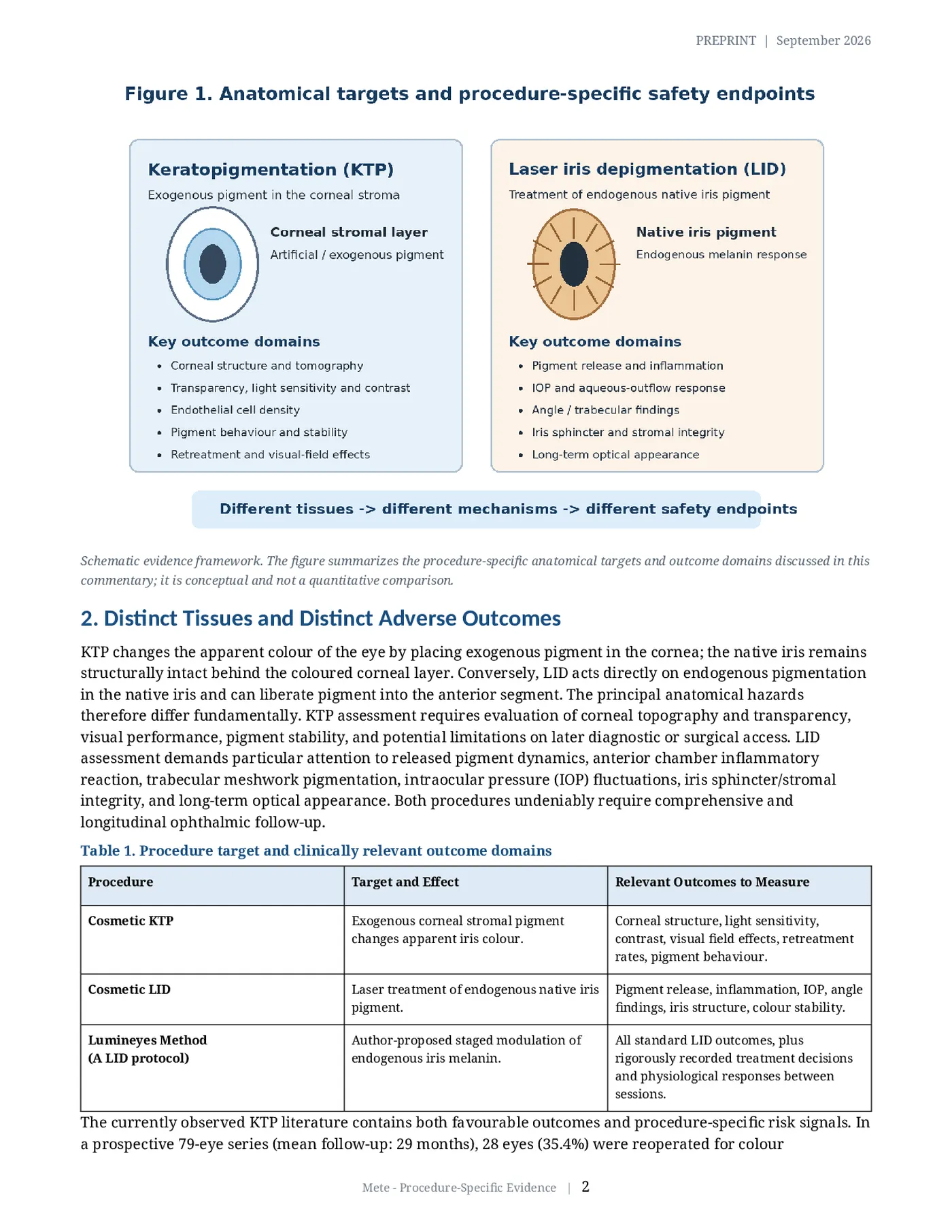
Schematic evidence framework. The figure summarizes the procedure-specific anatomical targets and outcome domains discussed in this commentary; it is conceptual and not a quantitative comparison.
2. Distinct Tissues and Distinct Adverse Outcomes
KTP changes the apparent colour of the eye by placing exogenous pigment in the cornea; the native iris remains structurally intact behind the coloured corneal layer. Conversely, LID acts directly on endogenous pigmentation in the native iris and can liberate pigment into the anterior segment. The principal anatomical hazards therefore differ fundamentally. KTP assessment requires evaluation of corneal topography and transparency, visual performance, pigment stability, and potential limitations on later diagnostic or surgical access. LID assessment demands particular attention to released pigment dynamics, anterior chamber inflammatory reaction, trabecular meshwork pigmentation, intraocular pressure (IOP) fluctuations, iris sphincter/stromal integrity, and long-term optical appearance. Both procedures undeniably require comprehensive and longitudinal ophthalmic follow-up.
Table 1. Procedure target and clinically relevant outcome domains Procedure Target and Effect Relevant Outcomes to Measure Cosmetic KTP Exogenous corneal stromal pigment Corneal structure, light sensitivity, changes apparent iris colour. contrast, visual field effects, retreatment rates, pigment behaviour.
Cosmetic LID Laser treatment of endogenous native iris Pigment release, inflammation, IOP, angle pigment. findings, iris structure, colour stability.
Lumineyes Method Author-proposed staged modulation of All standard LID outcomes, plus (A LID protocol) endogenous iris melanin. rigorously recorded treatment decisions and physiological responses between sessions.
The currently observed KTP literature contains both favourable outcomes and procedure-specific risk signals. In a prospective 79-eye series (mean follow-up: 29 months), 28 eyes (35.4%) were reoperated for colour adjustment, and light sensitivity at one month was reported in 30% [2]. A separate 166-eye prospective series reported no significant 12-month change in visual acuity, intraocular pressure, or visual field, but measured statistically significant decreases in endothelial cell density and specific contrast-sensitivity measures [3].
Furthermore, a five-patient case series described serious corneal ectatic changes following cosmetic KTP; while this identifies a critical possible complication, it supplies no definitive population incidence [4]. These findings collectively support the necessity of careful patient selection and targeted, procedure-specific follow-up without strictly negating the reported aesthetic benefits of KTP.
3. The Biological Fate of Pigment Matters
The author's earlier Research Archive emphasizes the central pathophysiological distinction as the persistence of introduced exogenous corneal pigment versus the alteration and potential biological processing of endogenous pigment native to the iris [7]. That framework serves as a mechanistic foundation, not a measured empirical claim that either route is inherently safer. KTP inherently requires rigorous attention to where deposited particles remain, whether they undergo microscopic migration or change appearance over decades, and how the surrounding corneal stroma responds chronologically. Conversely, in LID, liberated iris melanin material may remain in situ, redistribute, or enter anterior-segment clearance pathways (primarily the trabecular meshwork); complete, benign clearance cannot be universally assumed. While experimental rabbit iris models have reported melanophage responses following laser exposure [8], this does not directly establish the rate, efficiency, or safety of pigment clearance in treated human eyes. The cumulative amount, anatomical location, and duration of the pigment burden, combined with secondary inflammatory and aqueous outflow responses, require meticulous longitudinal observation.
The same Research Archive characterizes the Lumineyes method through three linked, author-proposed operational terms: selective stromal melanin modulation, controlled treatment-related pigment response, and response-guided staged treatment [7]. The term "selective" denotes the intended laser-tissue interaction but does not inherently prove absolute cellular specificity; "controlled" describes a continuously monitored clinical objective but cannot entirely exclude the possibility of serious pigment-related adverse events. Importantly, staging becomes a valid biological decision rule only when an observed physiological response actively dictates whether and how the subsequent exposure occurs. These standardized definitions significantly improve protocol reporting transparency, even while leaving definitive comparative effectiveness and overall safety unresolved pending larger trials.
4. Interpreting Reported Adverse Events
A previously published case following four cosmetic iris laser sessions documented severe bilateral intraocular pressures of 50 and 42 mmHg, extensive anterior-chamber pigment dispersion, and subsequent requirement for glaucoma surgery with persistent visual consequences [5]. This provides direct, undeniable evidence of severe potential harm and warrants explicit clinical attention during informed consent. However, a single case -lacking a defined treatment denominator or a fully comparable laser protocol description - cannot supply an accurate epidemiological event rate for all distinct forms of LID. Conversely, a separate series of 1176 eyes treated with photoablative cosmetic iridoplasty demonstrates that systematic, large-scale outcome reporting is achievable; yet, its specific modality and protocol should not be pooled uncritically with other distinct laser methods [6].
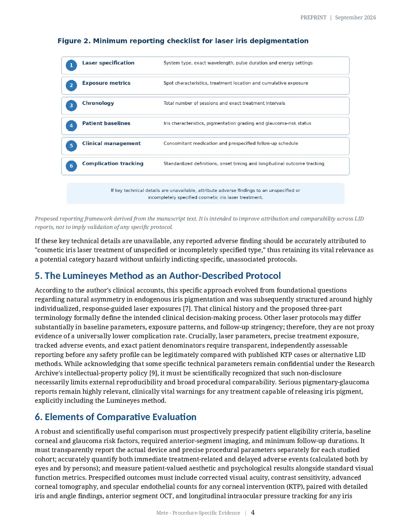
Proposed reporting framework derived from the manuscript text. It is intended to improve attribution and comparability across LID reports, not to imply validation of any specific protocol.
If these key technical details are unavailable, any reported adverse finding should be accurately attributed to "cosmetic iris laser treatment of unspecified or incompletely specified type," thus retaining its vital relevance as a potential category hazard without unfairly indicting specific, unassociated protocols.
5. The Lumineyes Method as an Author-Described Protocol
According to the author's clinical accounts, this specific approach evolved from foundational questions regarding natural asymmetry in endogenous iris pigmentation and was subsequently structured around highly individualized, response-guided laser exposures [7]. That clinical history and the proposed three-part terminology formally define the intended clinical decision-making process. Other laser protocols may differ substantially in baseline parameters, exposure patterns, and follow-up stringency; therefore, they are not proxy evidence of a universally lower complication rate. Crucially, laser parameters, precise treatment exposure, tracked adverse events, and exact patient denominators require transparent, independently assessable reporting before any safety profile can be legitimately compared with published KTP cases or alternative LID methods. While acknowledging that some specific technical parameters remain confidential under the Research Archive's intellectual-property policy [9], it must be scientifically recognized that such non-disclosure necessarily limits external reproducibility and broad procedural comparability. Serious pigmentary-glaucoma reports remain highly relevant, clinically vital warnings for any treatment capable of releasing iris pigment, explicitly including the Lumineyes method.
6. Elements of Comparative Evaluation
A robust and scientifically useful comparison must prospectively prespecify patient eligibility criteria, baseline corneal and glaucoma risk factors, required anterior-segment imaging, and minimum follow-up durations. It must transparently report the actual device and precise procedural parameters separately for each studied cohort; accurately quantify both immediate treatment-related and delayed adverse events (calculated both by eyes and by persons); and measure patient-valued aesthetic and psychological results alongside standard visual function metrics. Prespecified outcomes must include corrected visual acuity, contrast sensitivity, advanced corneal tomography, and specular endothelial counts for any corneal intervention (KTP), paired with detailed iris and angle findings, anterior segment OCT, and longitudinal intraocular pressure tracking for any iris intervention (LID). Importantly, an unobserved complication in a small cohort is not definitive proof of its absolute absence; 95% confidence intervals and detailed tracking of loss-to-follow-up must rigorously accompany all reported event counts. Distinct anatomical and physiological adverse outcomes should always be reported separately and transparently before drawing any overarching comparative conclusion.
Conclusion KTP and LID fundamentally target different ocular tissues and therefore categorically require distinct, highly specific outcome measures. Adverse events should strictly be attributed to fully documented protocols rather than broad categories. Establishing true comparative safety - explicitly including the evaluation of the Lumineyes method - mandates the use of comparable patient cohorts, adequate and standardized longitudinal follow-up, and ultimately, independent, peer-reviewed assessment.
Declarations
Article type Commentary/preprint; not peer reviewed.
Provenance The author's existing online Research Archive [7,9] supplied the conceptual framing; it is an author-published source, not independent clinical validation.
Ethics No original human or animal research or identifiable patient data.
Data availability No original data generated or analysed.
Funding No specific external funding.
Competing interest Mustafa Mete developed the Lumineyes method and is affiliated with MyLumineyes; he has a professional interest in the topic.
Related clinical background Laser Eye Color Change: https://mylumineyes.com/laser-eye-color-change/ (author website; provided for clinical context, not independent validation).
References
- D'Oria F, Alio JL. Surgical techniques for cosmetic eye color change: a narrative review. Ophthalmol Ther. 2025. doi:10.1007/s40123-025-01177-0.
- D'Oria F, Alio JL, Rodriguez AE, Amesty MA, Abu-Mustafa SK. Cosmetic keratopigmentation in sighted eyes: medium- and long-term clinical evaluation. Cornea. 2021;40:327-333. doi:10.1097/ICO.0000000000002417.
- Alio J, Sanginabadi A, Hojabr AT, Jafari B. Femtosecond laser-assisted keratopigmentation outcomes for pure cosmetic purposes. Am J Ophthalmol Case Rep. 2025;38:102297. doi:10.1016/j.ajoc.2025.102297.
- Alio JL, et al. Corneal ectasia following cosmetic keratopigmentation. Cornea. 2025. doi:10.1097/ICO.0000000000003754.
- Liu J, Korban S, Moster MR, Rhéaume MA, Wang Q. Bilateral severe iatrogenic pigmentary glaucoma following laser treatment for cosmetic iris color change. Am J Ophthalmol Case Rep. 2023;32:101927. doi:10.1016/j.ajoc.2023.101927.
- Grimaldos RP. Photoablative cosmetic iridoplasty: effective, safe, and predictable eye color change in 1176 eyes. Int Ophthalmol. 2021;41:1381-1393. doi:10.1007/s10792-021-01693-5.
- Mete M. Biological fate of pigment in cosmetic iris color modification: endogenous melanin modulation versus exogenous pigment deposition. MyLumineyes Research Archive. 2026. https://mylumineyes.com/research/research-biological-fate-pigment-iris-color-modification/ (accessed 28 September 2026). Author-published, not peer reviewed.
- Yildirim Y, et al. Evaluation of color-changing effect and complications after Nd:YAG laser application on iris surface. Med Sci Monit. 2016;22:107-114. doi:10.12659/MSM.895086.
- Mete M. MyLumineyes Research Library. 2026. https://mylumineyes.com/research/research-library/ (accessed 28 September 2026). Research index, not independent validation.
Original figures and table from the preprint
These are the exact source PDF pages containing the original figures/table; they are not newly generated clinical comparisons.