Risk Topology in Elective Eye-Color Modification: A Compartment-Based Framework for Corneal Keratopigmentation and Staged Laser Iris Depigmentation
Original source PDF (complete 12-page manuscript) · Zenodo DOI: 10.5281/zenodo.23458833
Author-hosted full text of the 2026 preprint. KTP means corneal keratopigmentation, not a KTP laser. The PDF remains authoritative for the original figure and layout.
Abstract
Background — Procedures that alter apparent eye color are frequently compared by their visible aesthetic result, although they impose perturbations in different ocular compartments. Contemporary keratopigmentation (KTP) introduces an exogenous pigment into the corneal stroma; laser iris depigmentation (LID) modifies endogenous iris pigmentation and may expose the anterior chamber to treatment-related pigment and inflammatory products. A useful comparison must therefore move beyond an undifferentiated ranking of procedures.
Objective — To develop a compartment-based framework that maps each intervention to its principal tissue exposure, temporal response and appropriate clinical surveillance domains.
Approach — A focused interpretation of published KTP outcome studies, experimental and clinical LID reports, complication cases, and reviews was combined with anatomical and optical reasoning. This article advances a conceptual framework and hypotheses; it does not pool studies or analyze new patient-level data.
Synthesis — KTP primarily creates a corneal tissue–material interface with documented postoperative ectasia, ulceration, photic symptoms, selected optical changes and pigment instability across non-equivalent clinical populations. LID creates an iris–aqueous interface in which pigment release, inflammation and intraocular-pressure (IOP) kinetics require explicit measurement. Published KTP studies offer a more developed clinical evidence base; LID evidence remains protocol-dependent and limited, with divergent outcomes across reports.
Neither anatomical difference nor unequal publication volume establishes comparative clinical superiority.
Conclusion — A multidimensional safety and function profile must include the clinically reported corneal complications, not solely eye-color change or corrected visual acuity. Compartment-specific surveillance, paired with a common core of functional outcomes, would make future studies more interpretable and allow hypotheses about each procedure to be tested independently.
Keywords: keratopigmentation; laser iris depigmentation; elective ophthalmology; corneal optics; pigment dispersion;
intraocular pressure; comparative outcomes; anterior segment.
1. Clinical problem and conceptual premise
Elective eye-color interventions present a methodological challenge: the observable cosmetic outcome is nearly identical in purpose, but the treated biological substrate can be entirely different. For outcome evaluation, cosmetic similarity is a poor surrogate for mechanistic equivalence. A colored corneal annulus and an altered anterior iris can look similar to an external observer while generating non-overlapping distributions of tissue exposure. A comparison that begins and ends with corrected distance visual acuity (CDVA), patient satisfaction or photographs will inevitably underdescribe these differences [1–3].
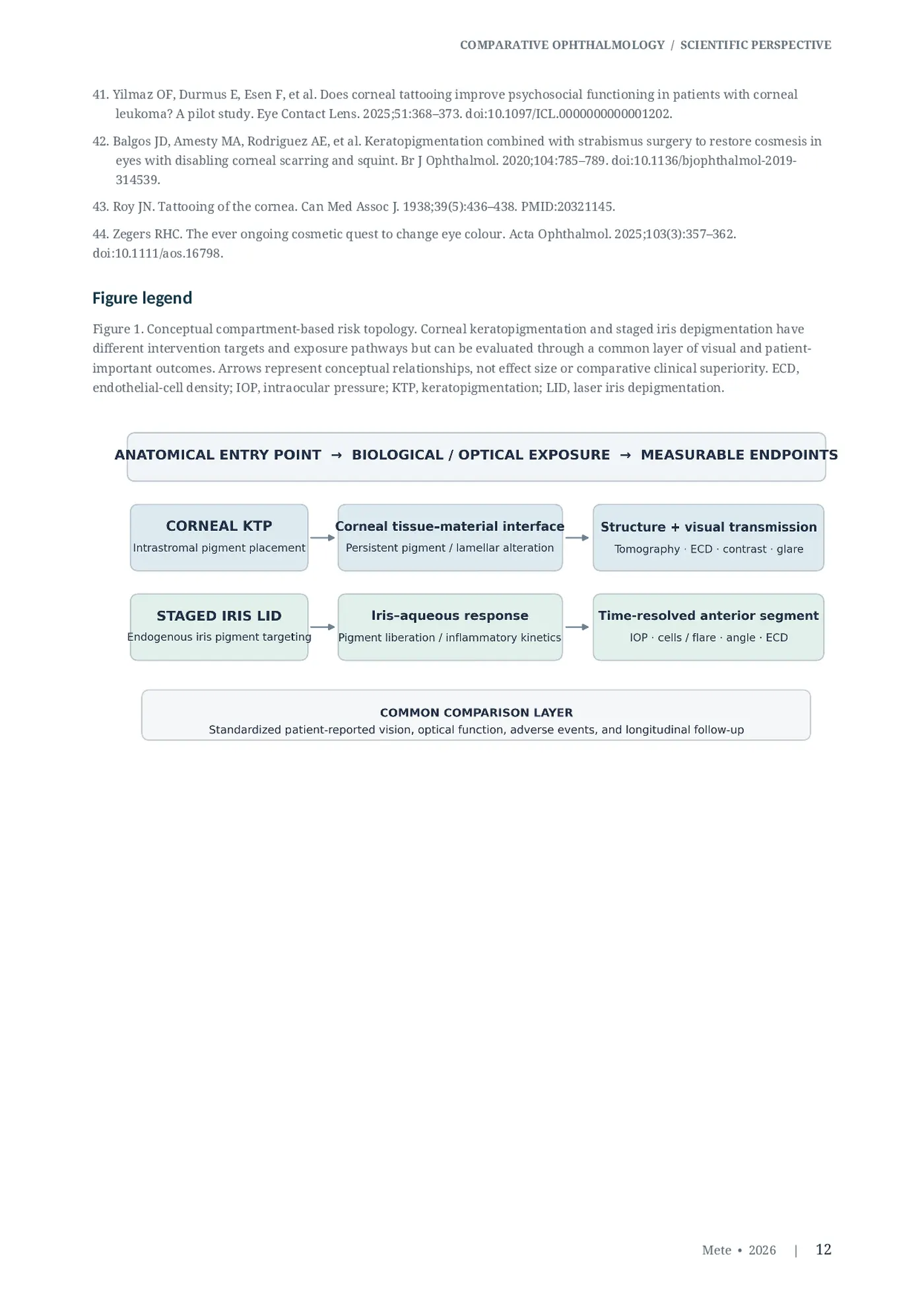
The relevant scientific question is not which procedure carries a universally larger “risk,” as though disparate adverse events could be placed on a single ordinal scale without empirical weighting. It is which structure is altered, what material is introduced or mobilized, how the induced response evolves over time, which functions are exposed, and what observations would discriminate benign adaptation from clinically important injury. This paper calls that organization a compartment-based risk topology: an anatomical map of plausible exposure pathways and the measurements needed to evaluate them.
Here, corneal KTP refers principally to contemporary pigment placement within a corneal stromal plane in sighted eyes. Staged LID refers to a category of approaches in which visible iris pigmentation is addressed over more than one treatment period with clinical response considered between exposures.
Different LID protocols are not assumed to be interchangeable. The discussion does not encompass artificial cosmetic iris implants, which constitute a separate intraocular category with an established complication literature [1,4]. 1.1 Historical evolution of corneal tattooing: from therapeutic camouflage to elective cosmetic pigmentation The concept of altering the apparent ocular surface through applied pigment substantially predates contemporary keratopigmentation. Historical accounts attribute the camouflage of conspicuous corneal leukomas to Galen in the second century CE and describe subsequent related practices in Byzantine medical writings associated with Aëtius of Amida. These early methods combined surface preparation or cauterization with topical pigment application; they were intended to conceal opacities in structurally compromised eyes, not to recolor the irides of otherwise healthy sighted individuals [2,43,44].
A major technical transition occurred in 1869, when Louis von Wecker described introducing dark pigment into corneal tissue by needle puncture. Corneal tattooing subsequently developed through different staining materials and manually created stromal entry methods, and continued to serve selected reconstructive indications, including visually conspicuous scars and the management of glare or photophobia related to iris defects. Twentieth-century clinical documentation and later therapeutic series establish continuity of the underlying principle of corneal coloration across changing instruments and indications [18,43,44].
Contemporary keratopigmentation introduced refined intrastromal pigment-delivery approaches, including lamellar dissection and laser-created stromal planes, along with purpose-developed pigment formulations. More recently, application has expanded from therapeutic camouflage or symptomatic rehabilitation to elective alteration of apparent eye color in healthy sighted eyes [2,4,28,44]. This is a clinically consequential shift in indication and risk tolerance rather than the invention of corneal pigmentation itself. Technical refinements may improve procedural control; their introduction alone does not establish long-term corneal biomechanical stability, pigment persistence or visual safety in healthy eyes. Historical continuity and modern clinical validation should therefore be treated as distinct questions.
2. Analytical approach and evidence boundaries
This is a mechanistic Scientific Perspective informed by selected peer-reviewed clinical series, experimental studies, case reports and contemporary reviews available by October 2026 [1–11,13–44].
The literature is interpreted at the level of procedure and study design, not pooled into a pseudo-comparative effect estimate. No prospective protocol, systematic eligibility screening, meta-analysis, new patient cohort or patient-level statistical analysis is claimed.
Three evidentiary levels are maintained throughout: (i) anatomical properties inherent to the intervention, such as a corneal pigment deposit in intrastromal KTP; (ii) measured outcomes in particular cohorts or reported cases; and (iii) mechanistic predictions that require prospective testing. This distinction is especially necessary when comparing the relatively mature cosmetic KTP literature with heterogeneous LID evidence. An observation made under one combination of laser, patient-selection and aftercare conditions should not be assigned automatically to a different protocol.
3. Compartment-based risk topology
3.1 Corneal tissue–material interface in KTP In intrastromal KTP, procedural exposure comprises the creation of a corneal plane and the retention of exogenous pigment within it. The resulting interface can be considered across four linked domains: tissue integrity, pigment–host interaction, optical transmission and long-term material behavior. Stromal lamellar manipulation provides a reason to assess tomography and biomechanics; retained pigment provides a reason to study color stability, inflammation and the spatial properties of transmitted light.
These are mechanistic obligations, not assertions that clinically important damage occurs in every treated eye [2,3,5,6].
The central clear aperture often remains optically functional, but it is spatially fixed relative to a pupil that changes size with illumination. Therefore, a normal photopic visual-acuity result does not logically exclude an effect on mesopic contrast, glare or peripheral rays. The size, eccentricity and optical density of the corneal pigment zone may all matter. These relations should be measured using standardized pupil size and viewing conditions rather than inferred from an image of eye color. 3.2 Iris–aqueous interface in LID LID does not require corneal pigment implantation or a stromal corneal tunnel. Its principal exposure is instead situated in the pigmented anterior iris and adjacent anterior-segment environment. Treatment can alter endogenous pigment distribution, with possible liberation of melanin-containing particles and an inflammatory response that interacts with aqueous outflow. In this setting, post-exposure kinetics are as relevant as the end-state iris appearance. Measurements restricted to a distant follow-up visit may fail to characterize short-lived IOP elevation, transient anterior-chamber cells or early corneal responses [7– 9].
There is an additional distinction within LID itself. Staged, response-guided protocols treat the interval between exposures as an opportunity for biological observation; other reported treatments may differ materially in laser characteristics, treated area, cumulative exposure and follow-up. The proposition that staging may attenuate peak biological burden is plausible but remains conditional on actual particle generation, clearance and individual outflow reserve. It cannot be demonstrated merely by naming a treatment “staged.” 3.3 Spatial and temporal non-equivalence KTP and LID differ simultaneously in where they act and how potential consequences may unfold. KTP introduces material and modifies corneal lamellar architecture, creating questions that can remain relevant over years. LID can produce an episodic response that may peak within a comparatively narrow observation window, while long-term iris structure and outflow health also remain clinically relevant.
The two procedures therefore require both common follow-up milestones and distinct early or late measurement emphasis. A lack of early events in a late-only dataset should not be taken as evidence that none occurred; equally, an isolated complication report cannot provide a population event rate.
4. Clinical evidence anchoring the framework
The 2021 prospective cosmetic KTP series of 79 eyes in 40 patients reported high aesthetic satisfaction at a mean follow-up of 29 months; 28 eyes (35.4%) underwent reoperation for cosmetic refinement. Light sensitivity, color evolution and a visual-field complaint were also recorded, while the series included an ectasia case in a patient with prior laser in situ keratomileusis [3]. The clinically relevant message is that a successful aesthetic outcome can coexist with retreatment demand and with uncommon but consequential structural or optical endpoints.
In a 2025 prospective 166-eye cosmetic KTP series, group-level CDVA, IOP, visual-field mean deviation and reported higher-order aberration measures did not show significant deterioration at 12 months. By contrast, mean endothelial-cell density decreased from 2393.29 to 2308.58 cells/mm², and contrast sensitivity declined across tested mesopic spatial frequencies, although investigators reported values remaining within normal limits [5]. These results support a differentiated rather than uniformly adverse interpretation of modern KTP: preservation of one metric does not render the other metrics redundant.
Separately, a five-patient, ten-eye case series documented progressive postoperative ectatic changes after cosmetic KTP, establishing a surveillance concern without establishing incidence in an unselected population [6].
The LID evidence base has a different structure. In the published 1,176-eye photoablative cosmetic iridoplasty series, the specific studied protocol was associated with favorable reported clinical outcomes, whereas a 2016 pigmented-rabbit experiment showed measurable iris depigmentation with short-lived low-grade inflammation under its experimental conditions [7,8]. Conversely, a 2023 report described severe bilateral pigmentary glaucoma with paracentral acute middle maculopathy following cosmetic iris laser sessions [9]. The latter event cannot be erased by a favorable series; nor can it be assigned as a measured complication rate for every technically distinct LID method. Together, the reports justify rigorous procedure-level descriptors and time-resolved physiological surveillance.
Reviews published in 2022 and 2025–2026 generally characterize the KTP evidence base as more extensive than that of LID while also identifying limitations in study size, duration, single-center concentration and long-term inference [1,4,10,11]. Evidence quantity, mechanistic properties and safety estimates must be evaluated separately.
5. A comparison architecture that does not collapse unlike outcomes
Comparative research requires a common core of patient-important outcomes plus dedicated measurements for each exposure compartment. The common core makes trials comparable; the specific modules prevent mechanistically important events from disappearing within a global summary score.
The table proposes an assessment architecture, not a validated risk index or a clinical rule for proceeding with treatment.
Table 1. Exposure-specific and common outcome domains Domain / purpose Corneal KTP: principal observation Staged LID: principal observation Tissue structure Corneal tomography, pachymetry, Iris morphology, anterior-segment epithelial and stromal morphology OCT, angle configuration Optical function Standardized mesopic contrast, glare Standardized mesopic contrast, glare and aperture–pupil interaction and pupil / iris behavior Cellular integrity Serial specular microscopy and Serial specular microscopy and endothelial-cell density endothelial-cell density Early biological response Ocular-surface and corneal Anterior-chamber cells / flare, visible inflammation, wound-related signs pigment, serial IOP Longitudinal response Ectatic change, pigment stability, Pigment / color stability, angle status, optical symptoms, retreatments IOP course, repeat exposures Shared patient outcomes CDVA, refraction, symptoms, quality of CDVA, refraction, symptoms, quality of vision, adverse events vision, adverse events Endpoint harmonization should define the eye and participant as distinct analytic levels, because bilaterally treated eyes are not statistically independent. Planned subgroup variables include baseline corneal status, previous refractive surgery, endothelial reserve, iris phenotype, baseline IOP and angle findings. Postoperative events require prespecified definitions and masked adjudication when possible. A large number of treated eyes should not be conflated with a correspondingly large number of independent patients.
6. A temporally resolved comparative research design
A clinically interpretable observational registry would prospectively enroll consecutive, suitably characterized individuals undergoing either approach, record explicit procedural exposure categories and report reasons for exclusion. It would measure the same baseline and long-term outcomes across groups while adding compartment-specific observations around relevant exposure windows. Matching or statistical adjustment would address preoperative ocular differences, but residual confounding should be expected in nonrandomized elective-treatment populations.
Baseline assessment should document CDVA, refraction, pupil behavior under defined luminance, photopic and mesopic contrast sensitivity, standardized color photography, ocular symptoms, corneal tomography, specular microscopy, IOP, gonioscopy and anterior-segment imaging. Immediately after treatment, the protocol should emphasize clinically relevant early response windows; for LID this includes serial pressure and anterior-chamber assessments rather than an isolated next-day value. At 1, 3, 6 and 12 months, and annually thereafter, the core battery should be repeated where feasible, with KTP-specific attention to topographic evolution and LID-specific attention to pressure and pigment-response history. These intervals are design suggestions, not empirically validated universal monitoring thresholds.
Safety analyses should describe denominator completeness, timing of observations, loss to follow-up and management of missing results. Retreatment must be analyzed both as a clinical intervention and as a patient-important outcome. Repeated LID sessions and KTP color touch-ups each introduce additional exposure, but their cumulative biological meaning differs and should not be equated by simple session counts. Standardized colorimetric outcomes should use controlled illumination, camera geometry and calibrated or reference-standard measurement, minimizing the influence of photographic conditions.
An event-specific safety table is preferable to a single binary “complication” endpoint. For example, clinically important ectasia, sustained visual-function loss, endothelial injury, severe IOP elevation and retinal ischemic sequelae require their own definitions and adjudication pathways. Event rates should be reported with appropriate confidence intervals; failure to observe a rare event in a small cohort does not establish its absence. 6A. Adverse-event spectrum: an evidence-stratified KTP literature extension An elective intervention in an otherwise healthy cornea must be judged by its capacity to preserve tissue as well as by cosmetic appearance. The evidence relevant to failure modes is heterogeneous: modern femtosecond cosmetic KTP in sighted eyes, therapeutic KTP in diseased or blind eyes, older manual corneal tattooing, pigment-tolerance experiments, and reviews do not supply interchangeable event denominators. The distinction is maintained in the following appraisal. The additional studies considered here extend the core literature substantially and are used to interrogate distinct risks rather than to inflate a pooled complication estimate [13–42]. 6A.1 Clinical ectasia and corneal biomechanical vulnerability Nouzovská and colleagues reported progressive topographic or refractive changes following cosmetic femtosecond-assisted KTP in 10 eyes of five patients, beginning 6 months to 4 years after surgery. Three underwent corneal cross-linking. Some eyes had additional susceptibility factors, including prior LASIK or persistent eye rubbing, whereas others had no clearly identified preoperative abnormality; one eye showed altered keratometry rather than unequivocal ectasia [6]. This is direct sighted-eye KTP evidence for a clinically relevant biomechanical signal, not an incidence estimate. The 2021 series of 79 eyes had previously recorded ectasia after KTP in a patient with a history of LASIK [3]. In a separate dysphotopsia series, irregular steepening and nonprogressive thinning followed repeat lamellar pigmentation in one eye; this evidence relates to a different indication, pigment and surgical technique [20].
These observations suggest a plausible interaction among lamellar geometry, pretreatment corneal reserve, rubbing or ocular allergy, prior refractive surgery and accumulated tissue intervention.
Femtosecond precision cannot logically eliminate a biological response to stromal dissection; conversely, an ectatic case after KTP cannot prove that the stromal pocket was the only causal contributor.
Prospective surveillance should therefore report baseline tomography, total pachymetric distribution, biomechanical variables when available, prior keratorefractive procedures, atopy, eye rubbing, retouches and serial posterior elevation. Longitudinal topographic change, not only postoperative central corneal thickness, should define the structural endpoint. 6A.2 Ulceration, microbial keratitis, delayed epithelial closure and corneal melting A 2025 ophthalmic case report from Uddin and colleagues documented severe corneal ulceration after purely cosmetic KTP, managed with amniotic membrane grafting and anti-inflammatory medication [13].
This is particularly important because it was observed in the elective cosmetic setting, although a single case cannot quantify frequency. In contrast, Hashemian and colleagues studied 85 eyes receiving therapeutic KTP for corneal scarring or iris abnormalities and described delayed epithelial healing (2.3%) and microbial keratitis (1.1%), along with dry eye, postoperative pain and photophobia [23]. In a 184-eye therapeutic blind-eye series, the reported postoperative spectrum included infectious keratitis, corneal melting and photophobia [16]. The latter two studies establish that tissue breakdown and infection belong to the clinical safety map, but their pre-existing corneal pathology prevents direct transfer of their rates to healthy elective eyes.
A large historical corneal tattooing cohort of 303 selected eyes, published online in 2026 as an article in press, reported persistent epithelial defects in 5.9%, microbial keratitis in 0.7% and corneal perforation in 1.0%. The underlying eyes were predominantly being rehabilitated for disfiguring ocular disease, and the techniques and pigments spanned several decades; the authors also found substantial differences between platinum chloride and India ink [15]. These observations are informative about material and wound vulnerability in compromised corneas, not about the incidence of perforation after contemporary femtosecond cosmetic KTP.
Older manual series reinforce the need for technique-specific interpretation. Rocher and colleagues described one perforation 40 days after tattooing in a markedly pathological cornea among 14 eyes [17]; Pitz and colleagues reported two inadvertent anterior-chamber perforations during manual tattooing of scarred, nonfunctional eyes [18]. Balgos and colleagues also reported corneal microperforation in a combined reconstructive cohort [42]. Describing any of these events as documented routine complications in previously healthy, femtosecond-treated cosmetic eyes would be inappropriate. Equally, excluding them altogether would obscure the clinical consequences of pigment delivery into injured corneal tissue and the importance of the underlying substrate. 6A.3 Photophobia, glare, aperture mismatch and subtle visual-function loss Postoperative photophobia is reproducible across distinct KTP evidence settings, though its magnitude and timing vary. Alio and colleagues, analyzing 234 eyes treated with several methods and pigment generations, characterized light sensitivity as the leading reported functional complaint; the frequently quoted 49% represents its share within the reported complication profile and must not be presented as a 49% rate among all 234 operated eyes [14]. In 79 sighted cosmetic eyes, light sensitivity occurred in 30% at one month, alongside color changes, fading and a visual-field limitation associated with a 4.5-mm aperture in one patient [3]. In 85 therapeutic eyes, photophobia was reported in 28.2% [23]. The distributions are not pooled because population and follow-up differ.
The 2025 prospective 166-eye cosmetic KTP study found no statistically significant group-level deterioration in visual acuity, visual-field mean deviation, IOP or reported higher-order aberrations at 12 months. Nevertheless, mean endothelial-cell density fell from 2393.29 to 2308.58 cells/mm² (approximately 3.5%), and mesopic contrast sensitivity decreased across the tested spatial frequencies, with selected photopic reductions; the authors reported contrast values within a normal range [5]. This apparently mixed result is not contradictory: high-contrast acuity and low-luminance contrast sensitivity interrogate different visual functions. Small or eccentric clear corneal apertures, pupil dilation and angular dependence of light transmission deserve separate testing rather than inference from CDVA or photographs [21,31,39]. 6A.4 Pigment fate: fading, chromatic change, neovascularization and retouch burden Pigment persistence does not imply pigment invariance. In the 234-eye mixed-indication series, fading, color change and neovascularization were reported, with outcomes varying across pigment generations [14]. The 79-eye cosmetic study documented reoperation in 35.4% of eyes, predominantly for color refinement [3]. In a 136-eye superficial therapeutic and cosmetic series, 44.7% of the first-stage group required one or more subsequent procedures, with postoperative complications among the subset followed for more than three months [24]. A separate 2019 study of 19 eyes treated for dysphotopsia after peripheral iridotomy found late depigmentation in 55% of the lamellar-pocket group after 3–5 years; commercial black ink and the functional indication limit extrapolation to newer mineral cosmetic pigments [20].
Calas and colleagues described three cases in which a previously implanted corneal tattoo changed to golden-brown five to six years after surgery; the authors discussed potential iron oxidation in the context of compromised corneas [19]. Laboratory investigations of micronized mineral pigments have evaluated corneal tissue tolerance and histopathological responses [25,26], while earlier clinical and longer follow-up reports document both acceptable pigment retention and later need for adjustment [27,28,30,32,40].
These strands should be viewed together: the question is not simply whether a pigment remains visible, but whether color, optical density, inflammatory behavior and local tissue response are stable under real ocular conditions. 6A.5 Optical access, microbiology, and the limits of biological generalization A 2023 study specifically tested ocular examinations six months after femtosecond-assisted cosmetic KTP in 60 eyes with a 5-mm central aperture. Investigators found routine multimodal screening feasible and interpretable, including assessment of the peripheral iris through translucent pigment, while peripheral endothelial cell counting was not possible [21]. It would therefore be erroneous to state that KTP universally blocks gonioscopy, retinal examination or future ophthalmic investigation. The narrower supported concern is examination- and region-specific attenuation of visibility that varies with pigment geometry, transparency, instrument and pathology. Longer follow-up and cross-center testing of posterior segment visibility under dilation remain worthwhile.
A 2024 prospective case-control investigation found no statistically significant alteration of conjunctival microbial culture patterns in patients with corneal tattoos compared with controls, and reported no infective complications in its institutional follow-up sample [22]. This is compatible with, not a refutation of, rare post-KTP microbial keratitis: changes in conjunctival colonization and risk of an exceptional wound-associated infection are different biological questions. Likewise, highly satisfactory results have been reported after KTP for corneal leukoma, iris defects or severe cosmetic disfigurement [23,27,29,33– 37,41]. Such therapeutic benefit cannot be dismissed, but neither does it establish the acceptable risk threshold for an entirely elective procedure on an optically healthy cornea.
One exceptional 2025 report described V1-distribution periocular skin hyperpigmentation after augmentation of a therapeutic corneal tattoo in an extensively operated eye [38]. The proposed mechanism of pigment migration is hypothetical, and a single unusual case provides no estimate of risk in contemporary elective KTP. A broader review of ocular and periocular tattoos identifies severe injuries across disparate tattoo techniques [39], but scleral and conjunctival-tattoo complications must not be counted as evidence of corneal KTP complications. The correct unit of attribution is the exact tissue, technique, pigment and clinical indication. 6A.6 Matrix of key reported harms and external-validity boundaries Table 3 separates direct modern cosmetic findings from therapeutic, historical and mechanistic evidence.
Its purpose is to preserve the seriousness of documented complications while preventing cross-population denominator errors.
Endpoint / evidence Population and observation Interpretive boundary Five cosmetic-KTP patients, 10 eyes; Direct sighted-eye signal; confounders Ectasia [6] postoperative progression 6 months–4 in some cases; no incidence years denominator Severe ulcer [13] One cosmetic-KTP eye; amniotic Direct cosmetic case; cannot estimate membrane graft required incidence 30% at 1 month in 79 cosmetic eyes; Timing, denominator and technique Photophobia [3,14,23] described also in mixed / therapeutic differ across studies cohorts 166 cosmetic eyes; 12-month mean Group mean; function remained Reduced contrast / ECD [5] ECD fall ~3.5% and mesopic contrast within tested normal ranges changes Keratitis / delayed healing [23] 85 therapeutic eyes; 1.1% microbial Underlying scars/iris defects; not a keratitis, 2.3% delayed healing cosmetic rate 184 blind eyes undergoing therapeutic High-risk substrate and multiple Corneal melt [16] pigmentation; melt included among techniques; no transfer to healthy eyes reported complications Older manual/therapeutic cohorts, No direct evidence of a comparable Perforation [15,17,18] including 1.0% in selected 303-eye perforation rate in modern cosmetic historic series KTP Iron-associated color change in 3 Pigment formulation and disease Late pigment instability [19,20] therapeutic eyes; late fading in status strongly modify external functional KTP validity 60 cosmetic eyes; routine screening Rejects universal examination-Diagnostic access [21] feasible, peripheral ECD assessment blockage claim; identifies spatial limited limitation Additional primary literature encompasses indications, technical variants and pigmentation longevity in older or diseased corneas [27–30,32–37,40–42]. These publications strengthen the assessment of external validity rather than creating additional head-to-head evidence against iris-based procedures. The empirical claim advanced here is deliberately specific: KTP has a documented, varied corneal safety profile that cannot be captured by satisfaction or acuity alone, while LID has a distinct pigment/aqueous/pressure profile requiring its own high-quality prospective characterization.
7. Falsifiable mechanistic hypotheses
The proposed topology generates testable questions that can be evaluated without disclosing proprietary laser parameters or assuming that a favored intervention is superior.
Table 2. Prospective hypotheses derived from anatomical exposure Hypothesis Mechanistic prediction Discriminating observation In KTP, optical symptoms may Paired pupil images, pigment-zone H1 — optical geometry correlate with pigment-annulus mapping, masked contrast and glare geometry relative to luminance- tests. dependent pupil size.
For LID, early IOP and inflammatory Repeated acute-window IOP and H2 — time dependence trajectories may carry information not anterior-chamber grades with captured by a single delayed prespecified time points. measurement.
Hypothesis Mechanistic prediction Discriminating observation KTP and LID will show different H3 — divergent structural endpoints distributions of corneal and iris Longitudinal tomography, AS-OCT and structural change despite a common endothelial imaging in both arms. cosmetic endpoint.
Stable high-contrast CDVA may coexist Within-person CDVA versus mesopic H4 — adequacy of conventional acuity with a meaningful change in other contrast, glare disability and patient-aspects of visual function. reported visual quality.
For H2, one plausible interpretation is that the temporal distribution of pigment liberation relative to aqueous clearance influences the height and duration of physiological response. However, particle load, clearance capacity and true tissue-level selectivity are not yet quantified sufficiently to infer safety from this relationship alone. The concept of response-guided selective stromal melanin modulation (SSMM) has been described separately as an author-proposed framework [12]; it is not treated here as an independently validated classification or as equivalent to the Grimaldos protocol.
8. Implications for interpretation and clinical reporting
The framework argues against two common inferential errors. The first is anatomical reductionism: claiming that avoidance of a corneal incision proves an iris-based method to be safer, despite an entirely separate anterior-chamber hazard domain. The second is evidentiary reductionism: treating a larger number of published KTP cases as sufficient to resolve every optical and structural endpoint or to eliminate the need for long-term monitoring. Each claim exceeds the measurements that support it.
A rigorous report should identify the target compartment, the origin of the pigment involved, the intervention-specific exposure window, the exact outcome measured and the population in which it was observed. An absence of reported IOP elevation after KTP does not answer the corneal biomechanical question; absence of a corneal pigment deposit after LID does not answer the outflow question. Similarly, a case report demonstrates possible harm but ordinarily cannot establish frequency.
This distinction is especially important where commercial and clinical interests coexist. Comparisons should be anchored in anatomical facts and protocol-specific evidence, with a transparent account of what remains to be tested. The strongest available KTP outcomes and the most serious documented LID outcomes should both remain visible in the analysis, because excluding either would distort the research agenda.
9. Limitations
This analysis is an evidence-stratified mechanistic perspective, not a comprehensive systematic review.
Published KTP and LID cohorts are not directly comparable in patient selection, procedure definition, sample size, length of follow-up or measurement schedules. Published severe adverse-event reports are vulnerable to reporting bias, while favorable single-center series may be affected by selection and ascertainment bias. Some proposed endpoints have uncertain repeatability across instruments and clinics. None of the hypotheses in Table 2 has been established as a comparative causal mechanism by this article. The framework should be refined through independent measurement, preregistered endpoints and replication rather than adopted as a ranking system.
10. Conclusion
Corneal keratopigmentation and staged laser iris depigmentation must be compared as different biological perturbations, not merely alternative routes to the same visible color. KTP concentrates intervention-related questions in corneal structure, pigment retention and optical geometry; LID concentrates additional questions in the iris–aqueous interface and its time-dependent response. Their outcome profiles overlap at the level of patient visual function, but their mechanistic surveillance cannot be identical. A dual strategy—shared functional endpoints plus compartment-specific structural and physiological endpoints—offers a more discriminating basis for future comparative ophthalmic research than categorical claims based on cosmetic appearance alone.
Declarations
Funding: No external funding.
Competing interests: Mustafa Mete is the developer of Lumineyes™ and originator of the SSMM conceptual framework and has a professional and proprietary interest in laser iris depigmentation.
Data availability: No new patient-level data were collected or analyzed for this article. All cited published reports are identified in the references.
References
- D’Oria F, Alio JL. Surgical Techniques for Cosmetic Eye Color Change: A Narrative Review. Ophthalmol Ther. 2025;14:1685– 1694. doi:10.1007/s40123-025-01177-0.
- Hasani H, Es’haghi A, Rafatnia S, Alilou S, Abolmaali M. Keratopigmentation: a comprehensive review. Eye (Lond). 2020;34:1039–1046. doi:10.1038/s41433-019-0750-2.
- D’Oria F, Alio JL, Rodriguez AE, Amesty MA, Abu-Mustafa SK. Cosmetic Keratopigmentation in Sighted Eyes: Medium- and Long-term Clinical Evaluation. Cornea. 2021;40:327–333. doi:10.1097/ICO.0000000000002417.
- D’Oria F, Abu-Mustafa SK, Alio JL. Cosmetic Change of the Apparent Color of the Eye: A Review on Surgical Alternatives, Outcomes and Complications. Ophthalmol Ther. 2022;11:465–477. doi:10.1007/s40123-022-00458-2.
- Alio JL, Sanginabadi A, Hojabr AT, Jafari B. Femtosecond laser-assisted keratopigmentation outcomes for pure cosmetic purposes. Am J Ophthalmol Case Rep. 2025;38:102297. doi:10.1016/j.ajoc.2025.102297.
- Nouzovská K, Barazandeh Noveyri B, Abu-Mustafa SK, Alió JL. Corneal Ectasia Following Cosmetic Keratopigmentation. Cornea. 2025;44:779–787. doi:10.1097/ICO.0000000000003754.
- Grimaldos Ruiz P. Photoablative cosmetic iridoplasty: effective, safe, and predictable—eye color change in 1176 eyes. Int Ophthalmol. 2021;41:1381–1393. doi:10.1007/s10792-021-01693-5.
- Yildirim Y, Duzgun E, Kar T, et al. Evaluation of Color-Changing Effect and Complications After Nd:YAG Laser Application On Iris Surface. Med Sci Monit. 2016;22:107–114. doi:10.12659/MSM.895086.
- Liu J, Korban S, Moster MR, Rhéaume MA, Wang Q. Bilateral severe iatrogenic pigmentary glaucoma following laser treatment for cosmetic iris color change. Am J Ophthalmol Case Rep. 2023;32:101927. doi:10.1016/j.ajoc.2023.101927.
- Hashemi H, Hadavandkhani A, Khabazkhoob M. An updated comprehensive review on keratopigmentation. Int J Ophthalmol. 2025;18:2183–2194. doi:10.18240/ijo.2025.11.22.
- Alio JL. Cosmetic Keratopigmentation Works and Can Be Used as a New Aesthetic Solution to Change the Apparent Color of the Eye. Cornea. 2026;45:809–810. doi:10.1097/ICO.0000000000004172.
- Mete M. Selective Stromal Melanin Modulation (SSMM), version 1.5. Zenodo. 2026. doi:10.5281/zenodo.23424601.
- Uddin O, Shah Y, Majidi S, Mei F, Eghrari AO, Akpek E. Severe Corneal Ulceration After Cosmetic Keratopigmentation. Cornea. 2025;44:1319–1321. doi:10.1097/ICO.0000000000003895.
- Alio JL, Al-Shymali O, Amesty MA, Rodriguez AE. Keratopigmentation with micronised mineral pigments: complications and outcomes in a series of 234 eyes. Br J Ophthalmol. 2018;102:742–747. doi:10.1136/bjophthalmol-2017-310591.
- Alzaben K, Asiri A, Alaidan A, et al. Long-Term Outcomes of Corneal Tattooing for Cosmetic Rehabilitation of Disfigured Blind Eyes. AJO International. Available online 29 Aug 2026;100307 (article in press). doi:10.1016/j.ajoint.2026.100307.
- da Costa AX, Cal AFF, Bannach MP, de Oliveira GH, Garotti G. Therapeutic keratopigmentation Vis-à-Vis in blind eyes: complications and outcomes in a series of 184 eyes. Front Med (Lausanne). 2025;12:1700788. doi:10.3389/fmed.2025.1700788.
- Rocher N, Hirst L, Renard G, Doat M, Bourges JL, Mancel E. De l'usage du tatouage cornéen: à propos de 14 cas [Corneal tattooing: 14 cases]. J Fr Ophtalmol. 2008;31:968–974. doi:10.1016/S0181-5512(08)74742-9.
- Pitz S, Jahn R, Frisch L, Duis A, Pfeiffer N. Corneal tattooing: an alternative treatment for disfiguring corneal scars. Br J Ophthalmol. 2002;86:397–399. doi:10.1136/bjo.86.4.397.
- Calas E, Gueudry J, Muraine M. Severe color change in corneal tattoos: Report of 3 cases. J Fr Ophtalmol. 2018;41:e469– e475. doi:10.1016/j.jfo.2018.10.001.
- Jabbour S, Choremis J, Boutin T, et al. Poor Long-Term Outcomes of Keratopigmentation With Black Ink for the Treatment of Dysphotopsia Secondary to Laser Peripheral Iridotomies. Cornea. 2019;38:1400–1405. doi:10.1097/ICO.0000000000002117.
- Alafaleq M, Garrido J, Ferrari F. Femtosecond assisted keratopigmentation with a 5-millimeter diameter aperture does not affect ocular examination. Eur J Ophthalmol. 2023;33:1898–1902. doi:10.1177/11206721231158275.
- Yilmaz OF, Sarmis A, Mutlu MA, Oguz H. Does corneal tattooing affect the conjunctival microbiota? Cutan Ocul Toxicol. 2024;43:46–51. doi:10.1080/15569527.2023.2275025.
- Hashemian SJ, Roozdar S, Alió JL. Therapeutic Keratopigmentation: Cosmetic and Functional Outcomes. Ophthalmol Ther. 2025;14:1133–1145. doi:10.1007/s40123-025-01131-0.
- Al-Shymali O, Rodriguez AE, Amesty MA, Alio JL. Superficial Keratopigmentation: An Alternative Solution for Patients With Cosmetically or Functionally Impaired Eyes. Cornea. 2019;38:54–61. doi:10.1097/ICO.0000000000001753.
- Amesty MA, Alio JL, Rodriguez AE. Corneal tolerance to micronised mineral pigments for keratopigmentation. Br J Ophthalmol. 2014;98:1756–1760. doi:10.1136/bjophthalmol-2014-305611.
- Amesty MA, Rodriguez AE, Hernández E, et al. Tolerance of micronized mineral pigments for intrastromal keratopigmentation: a histopathology and immunopathology experimental study. Cornea. 2016;35:1199–1205. doi:10.1097/ICO.0000000000000900.
- Alio JL, Sirerol B, Walewska-Szafran A, et al. Corneal tattooing with new mineral micronised pigments to restore cosmetic appearance in severely impaired eyes. Br J Ophthalmol. 2010;94:245–249. doi:10.1136/bjo.2008.149435.
- Alió JL, Rodriguez AE, El Bahrawy M, et al. Keratopigmentation to change the apparent color of the human eye: a novel indication for corneal tattooing. Cornea. 2016;35:431–437. doi:10.1097/ICO.0000000000000745.
- Karslioglu MZ, Tas AY, Kesim C, Sahin A, Muftuoglu O. Keratopigmentation: Is It a Miracle or an Adventure? Beyoglu Eye J. 2020;5:32–37. doi:10.14744/bej.2020.76476.
- Yin HY, Walter KA. Long-Term Outcome of Femtosecond Laser-Assisted Keratopigmentation: Using Intacs Channels for Precise Pigment Deposition Within the Cornea. Cornea. 2021;40:1330–1335. doi:10.1097/ICO.0000000000002667.
- Alafaleq M, van Haselen R, Ferrari F. The safety and effectiveness of a novel annular keratopigmentation technique: a cross-sectional survey of patients. BMC Ophthalmol. 2023;23:292. doi:10.1186/s12886-023-02911-7.
- Yilmaz OF, Oguz H. Aesthetic corneal tattooing/keratopigmentation using tattoo pen machine: choosing suitable method and color. Graefes Arch Clin Exp Ophthalmol. 2023;261:3531–3540. doi:10.1007/s00417-023-06135-9.
- Mohan S, Tripathi A, Patel N, et al. Intrastromal keratopigmentation: a boon for unsightly corneal scars. Int Ophthalmol. 2023;43:2787–2794. doi:10.1007/s10792-023-02700-7.
- Trindade BLC, Coelho IB, Magalhães LL, et al. Cosmetic Therapeutic Keratopigmentation. Clin Ophthalmol. 2025;19:527– 534. doi:10.2147/OPTH.S507490.
- Alsmman AH, Mostafa EM, Mounir A, et al. Outcomes of corneal tattooing by Rotring painting ink in disfiguring corneal opacities. J Ophthalmol. 2018;2018:5971290. doi:10.1155/2018/5971290.
- Doganay D, Doganay S, Cankaya C. Corneal tattooing for esthetic purposes in patients with corneal opacities. Indian J Ophthalmol. 2020;68:1033–1036. doi:10.4103/ijo.IJO_1502_19.
- Agarwal R, Urkude J, Nair S, et al. Effect of intraoperative optical coherence tomography on anatomic and cosmetic results of intrastromal tattooing. Saudi J Ophthalmol. 2021;35:39–46. doi:10.4103/1319-4534.325784.
- Alsuliman N, Alruwaili S. Post-corneal tattooing trigeminal nerve-distributed hyperpigmentation: a case report. BMC Ophthalmol. 2025;25:526. doi:10.1186/s12886-025-04364-6.
- Lee KWA, Chan LKW, Lee AWK, Lee CH, Wan J, Yi KH. Ocular and Periocular Tattoo Adverse Effects: A Review. Diagnostics (Basel). 2024;14:2150. doi:10.3390/diagnostics14192150.
- Ferrari F, van Haselen R. The safety and effectiveness of a novel annular keratopigmentation method: a case report. Case Rep Ophthalmol. 2018;9:35–42. doi:10.1159/000485554.
- Yilmaz OF, Durmus E, Esen F, et al. Does corneal tattooing improve psychosocial functioning in patients with corneal leukoma? A pilot study. Eye Contact Lens. 2025;51:368–373. doi:10.1097/ICL.0000000000001202.
- Balgos JD, Amesty MA, Rodriguez AE, et al. Keratopigmentation combined with strabismus surgery to restore cosmesis in eyes with disabling corneal scarring and squint. Br J Ophthalmol. 2020;104:785–789. doi:10.1136/bjophthalmol-2019-314539.
- Roy JN. Tattooing of the cornea. Can Med Assoc J. 1938;39(5):436–438. PMID:20321145.
- Zegers RHC. The ever ongoing cosmetic quest to change eye colour. Acta Ophthalmol. 2025;103(3):357–362. doi:10.1111/aos.16798.
Figure legend
Figure 1. Conceptual compartment-based risk topology. Corneal keratopigmentation and staged iris depigmentation have different intervention targets and exposure pathways but can be evaluated through a common layer of visual and patient-important outcomes. Arrows represent conceptual relationships, not effect size or comparative clinical superiority. ECD, endothelial-cell density; IOP, intraocular pressure; KTP, keratopigmentation; LID, laser iris depigmentation.
Original manuscript figure
Source-page facsimile reproduced directly from the original 2026 preprint PDF; the complete PDF remains authoritative.