Selective Stromal Melanin Modulation (SSMM): an integrative biophysical and clinical framework for response-guided laser iris pigment modification
Original preprint date: 13 November 2026 · Archived DOI: 10.5281/zenodo.24203344
Status: author-hosted copy of the existing preprint; not a separate peer-reviewed journal publication.
Read or download the complete 19-page original PDF, including Tables 1–4 and Figures 1–3 · Zenodo archival record
Abstract
Purpose: To integrate ocular pigment biology, laser–tissue interaction, and longitudinal clinical assessment into a falsifiable framework—selective stromal melanin modulation (SSMM)—for studying laser-directed anterior iris pigment modification.
Methods: A targeted narrative review of relevant PubMed/MEDLINE-indexed and foundational literature, supplemented by reference tracking through 13 November 2026, was used to distinguish empirical observations, mechanistic analogies, and hypotheses. No new clinical cohort was analyzed. The proposed Target–Response–Decision architecture was evaluated against experimental iris research, published human laser studies, and reported complications.
Results: SSMM specifies biophysical, anatomical, and functional dimensions of selectivity, distinguishes pigment interaction from pigment processing and tissue response, and incorporates longitudinal response selectivity (LRS). Seven propositions are linked to candidate measurements, comparators, and disconfirming results. Published severe adverse outcomes, uncertainty in pigment load and outflow response, and the lack of protocol-specific endothelial and exposure data remain constraints on interpretation.
Conclusion: SSMM is a research framework, not an established selective or safe treatment mechanism. Its principal contribution is to make target specificity, stromal preservation, temporal differentiation, and stage-informed decision value independently testable.
Keywords: iris stroma; selective stromal melanin modulation; laser–tissue interaction; longitudinal response selectivity; laser iris depigmentation; anterior-segment imaging
Introduction
Human iris coloration is determined by the quantity and spatial organization of melanin, stromal architecture, optical absorption, and scattering. Morphological studies associate visible color differences more closely with the amount and distribution of stromal pigment than with melanocyte counts alone [1, 2]. The iris is also a functional tissue comprising melanocytes, fibroblasts, vascular and neural elements, and extracellular matrix; an intervention directed at pigment cannot be interpreted exclusively as a color intervention [3, 4].
The resulting research problem is to distinguish the intended chromophore interaction from concurrent changes in tissue integrity and ocular physiology. Visible color change is a phenotype, but it is not by itself evidence of selective target engagement, pigment clearance, or absence of injury. The principle of selective photothermolysis provides a starting point for assessing preferential interaction with an absorbing chromophore [5].
Optical behavior additionally depends on wavelength, pulse duration, delivered energy and fluence, beam geometry, optical scattering, and local tissue composition [6]. Photothermal and photomechanical pathways can coexist, and selectivity cannot be inferred from melanin absorption alone [7]. Published work includes controlled rabbit iris experiments [8], a human clinical series with staged photoablative iridoplasty [9], and broader reviews of cosmetic eye-color interventions [10, 11].
These sources differ in exposure, design, follow-up, and ascertainment of outcomes. Importantly, severe secondary pigmentary glaucoma has also been documented after cosmetic iris laser treatment [18, 27]. Shared cosmetic intent does not establish equivalence of protocols, nor does protocol heterogeneity invalidate documented serious complications. The term laser iris depigmentation describes a clinical aim or observed appearance but does not, by itself, define which iris compartments are exposed, the physical interaction, pigment processing, or the sequence of clinical decisions.
SSMM was proposed in the Lumineyes research program as an explicit integration of those questions [12].
Review approach, scope, and scientific background
Literature identification and selection. A targeted, non-systematic review was undertaken using PubMed/MEDLINE-indexed primary and review literature identified through 13 November 2026. ” Reference lists of relevant original studies and reviews were also examined. Sources were selected for direct relevance to iris morphology, optical interaction, anterior-segment response, clinical laser observations, or competing mechanisms.
Implant-based and corneal pigmentation techniques were considered only where needed to distinguish modalities. This was not a systematic search or meta-analysis, and no claim of exhaustive coverage or pooled event rates is made [30]. Evidence classification. Human histological and cellular studies establish anatomical or cellular facts; controlled experimental studies demonstrate responses only under their tested conditions; clinical series describe outcomes within their own sampling and ascertainment methods; and case reports establish the possibility of serious events but cannot estimate incidence.
Proposed SSMM constructs and the author’s earlier Zenodo records document conceptual provenance rather than validation. Each evidentiary level is kept separate in the synthesis. The Lumineyes™ method evolved from exploratory work initiated in 2009 and was further developed clinically during 2017–2026. This history explains the questions that motivated the SSMM construct; it is not presented here as a prospectively defined or independently audited participant cohort.
No patient-level outcomes, adverse-event frequencies, or protocol-specific comparative effects have been derived for the present article. The framework asks four linked questions: (i) can pigment-bearing stromal targets be preferentially affected under defined exposures; (ii) how does visible pigment change relate to structural and cellular response; (iii) do baseline phenotype and sequential observations improve interpretation; and (iv) can clinically important ocular responses be differentiated over time without assuming that color change implies tissue preservation?
The analysis proceeds from optical target and physical exposure to pigment processing, cellular and physiological effects, repeated measurement, and decision assessment. Questions of safety, efficacy, and causal mechanism are posed as empirical endpoints rather than presumed consequences of the model.
Operational Definition and Three Dimensions of Selectivity
Selective stromal melanin modulation is a proposed strategy for studying preferential optical interaction with melanin-bearing structures in the anterior iris stroma and the resulting pigmentary, anatomical, and physiological responses during staged observation. It does not imply that clinical stromal sparing, an exposure threshold, or long-term safety has been demonstrated. “Selective” refers to an experimentally testable preferential response, not exclusive absorption by melanin or proof of no collateral effect.
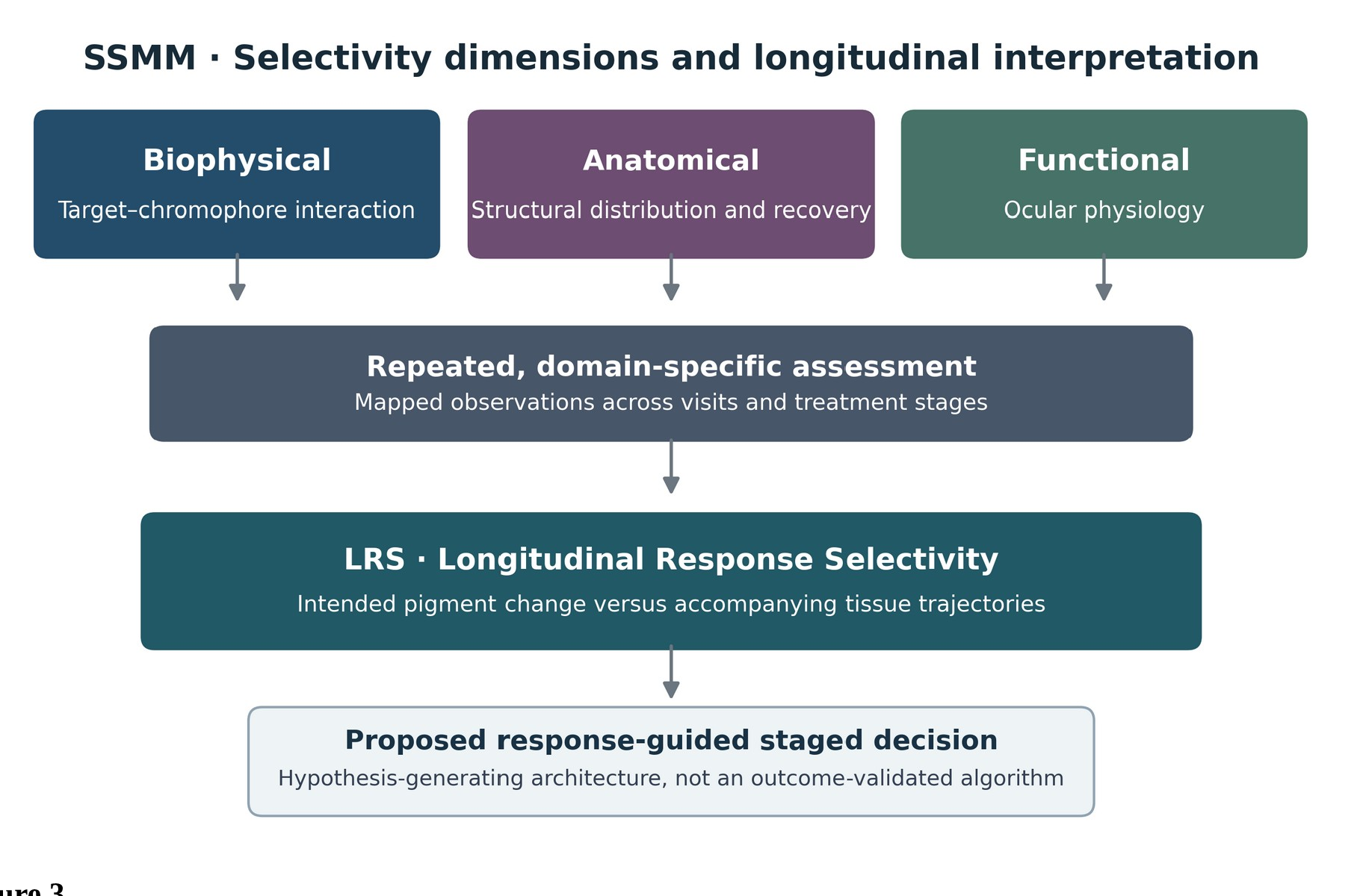
“Stromal” denotes the intended anatomical compartment; “modulation” distinguishes the research objective of pigment change from indiscriminate structural removal. The term is descriptive of a hypothesis and does not establish its clinical fulfillment. Three dimensions are operationally separated: biophysical selectivity asks whether target-associated absorption and physical response differ from suitable non-target comparators; anatomical selectivity asks whether secondary tissue alteration is spatially or quantitatively dissociable from pigment change; and functional selectivity examines ocular physiological outcomes after exposure.
Evidence in one dimension cannot substitute for evidence in either of the others (Table 1).
Biophysical Foundations of Stromal Pigment Modification
Melanin and the local optical environment
Melanin is an endogenous ocular chromophore, but its response to optical radiation is conditional on absorption spectrum, pulse duration, irradiance, scattering, pigment localization, and surrounding tissue [4– 7]. SSMM designates anterior stromal melanin-bearing structures as its intended region of study; it does not treat the iris as optically homogeneous. The same gross iris color may conceal distinct pigment depth, density, and spatial heterogeneity.
This suggests a testable phenotype–response relationship; appearance alone is an inadequate proxy for local optical dose or total released pigment.
Localized pigment interaction
Both photothermal and photomechanical pathways are plausible during short-pulse exposure of pigmented tissues [7]. Microcavitation, if present, would require experiment-specific evidence regarding thresholds, bubble dynamics, and nearby stromal and endothelial effects. An observed color change cannot discriminate between these interactions. Thermal confinement and stress confinement are different physical criteria, governed by pulse duration relative to local heat-diffusion and acoustic-transit times.
Work in retinal and melanosome models shows that pulse shape affects microcavitation behavior [13]; such thresholds should not be transferred directly to living human iris. Meaningful verification of preferential stromal response must specify wavelength, pulse width, spot profile, delivered fluence, repetition, cumulative exposure, target geometry, and appropriate structural readouts.
This conceptual review does not analyze a complete Lumineyes-specific exposure dataset and therefore makes no quantitative threshold or tissue-sparing claim.
Direct experimental iris evidence
In 12 pigmented rabbits, Yildirim et al. investigated 532-nm Q-switched Nd:YAG exposure and reported reduced anterior iris stromal pigmentation accompanied by pigment-bearing macrophages [8]. Within the reported conditions, they did not identify posterior iris pigment epithelial histological alterations. This supports the possibility of separable pigment alteration and cellular handling in that experimental model; it does not demonstrate clinical equivalence, quantify anterior-chamber pigment burden, or exclude effects under other exposure regimes.
Controlled Pigment Response: Modification, Processing, and Recovery
“Controlled pigment response” is used as a decision and measurement concept: intended exposure is followed by observation, assessment, and a subsequent clinical decision. It does not indicate that liberated pigment mass or the aqueous clearance capacity has been quantified. For clarity, three processes are separated analytically: initial pigment modification at the target; processing of liberated or redistributed pigment (including possible cellular uptake and outflow exposure); and later biological or anatomical tissue response.
The sequence is conceptual, not proof that these processes occur in fixed order or reach complete recovery. Pigmentary change, intracellular or extracellular pigment handling, and tissue response may proceed at different rates and may diverge between eyes or across stages. Colorimetric endpoints, aqueous particle measurements, optical imaging, and physiological measures therefore answer different questions and require distinct controls.
Visible lightening alone cannot quantify pigment mass, determine where pigment is deposited, or show that stromal matrix and melanocyte function remain intact. The proposed P-CLEAR construct distinguishes release and processing questions but is a research proposal, not evidence of successful clearance [14]. Human iris studies show that melanocyte pigmentation may be influenced by fibroblast-dependent signals [15] and that matrix metalloproteinases and their inhibitors are present in iris/ciliary tissues [16].
These are relevant biological properties, but neither paper tests post-laser stromal regeneration or recovery after pigment modification. The present synthesis uses them to identify distinct measurable cell–matrix domains, not to infer an equivalent laser-repair pathway.
Distinction From Classical Pigment Dispersion Syndrome
In classical pigment dispersion syndrome (PDS), pigment frequently originates from the posterior iris pigment epithelium in association with iris–zonular contact [17]. The SSMM target hypothesis concerns anterior stromal melanin. These are distinguishable starting compartments, but liberated pigment may converge on the aqueous and trabecular outflow pathways regardless of origin (Figure 1).
Source-specific morphology does not imply a safer downstream course. Particle number, size, aggregate chemistry, duration of exposure, inflammatory response, and the receiving outflow system must be examined before predicting trabecular effects. Pigment challenge experiments show altered trabecular meshwork phagocytosis, contraction, and migration under their tested conditions [28].
They do not supply a dose-response boundary for a clinical SSMM protocol. Documented severe complications after cosmetic iris laser exposure include refractory secondary pigmentary glaucoma requiring filtration surgery and, in one report, paracentral acute middle maculopathy associated with marked pressure elevation [18, 27]. These reports establish important counterexamples to any generic assumption that a cosmetic pigment endpoint is benign.
Their exposure details and denominators are insufficient to estimate protocol-specific event rates; dedicated standardized outcome studies are needed rather than extrapolation in either direction.
The Lumineyes™ Conceptual Fingerprint
The Lumineyes™ Conceptual Fingerprint organizes the research questions as Target → Response → Decision: intended pigment-bearing anatomy and exposure; optical, cellular, anatomical, and physiological effects; and whether sequential observations improve subsequent assessment. This is a structure for hypothesis testing, not a validated treatment algorithm. Staged clinical treatment and reassessment have already been reported in human photoablative iridoplasty [9].
SSMM does not claim to have originated staging itself. Its proposed added value is to keep three types of selectivity separate, specify the pigment-versus-tissue distinction, and test whether the information obtained between stages changes subsequent predictions. Selective laser trabeculoplasty (SLT) constitutes a mechanistically distinct intervention at the trabecular meshwork.
Alvarado et al. demonstrated monocyte-associated changes in outflow-related experiments after SLT [19]. This cannot establish the same response after anterior stromal pigment exposure: the target, particle burden, and sequence of injury differ. Conversely, pigment-challenge experiments indicate that excess pigment may impair trabecular cell functions [28]. These observations motivate competing hypotheses —obstruction or dysfunction versus adaptive cellular response—that must be resolved through direct outflow and pigment-burden measurements.
An IOP change alone would not identify the mechanism.
Phenotype and Interindividual Response Variability
Regional optical accessibility cannot be deduced reliably from a broad visible color category. Spatial heterogeneity and depth-specific pigment distribution are possible sources of between-eye variation and motivate pre-exposure imaging and standardized regional analysis. The proposed Lumineyes Iris Pigmentation Scale (LIPS) describes apparent anterior stromal pigmentation in five categories [20].
Grades I–IV are ordered by diffuse apparent pigmentation; Grade V is defined by marked spatial heterogeneity and should not be read as a darker grade above IV. LIPS is not yet a validated severity measure, and its interobserver reliability, sensitivity to change, and predictive utility remain empirical questions. Lumineyes Optical Architectural Typology (L-OAT) is a separate proposed framework for optical and architectural phenotype [21].
Neither naming a phenotype nor assigning a category demonstrates that it predicts response. Independent grading, standardized acquisition, and out-of-sample performance testing are required.
Longitudinal Response Selectivity (LRS)
Definition and position
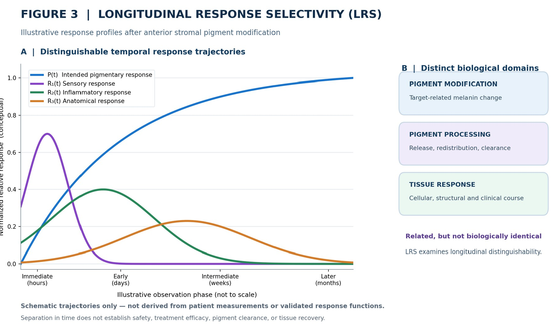
Longitudinal Response Selectivity (LRS), proposed within the SSMM framework in 2026, is defined below as an evaluation concept rather than an additional optical targeting mechanism. Longitudinal Response Selectivity (LRS) is the proposed extent to which intended stromal pigment modification can be distinguished from accompanying tissue responses across successive observation periods and treatment stages.
Operationally, LRS couples repeated observation with separate definitions for pigmentary and non-pigmentary responses (Figure 2). Distinguishable trajectories would not prove selective targeting or preserved anatomy; those questions require independent endpoints.
Temporal interpretation
The exposure is brief relative to the possible evolution of pigmentary, sensory, inflammatory, anatomical, and functional responses. Measurements restricted to one postoperative time point may therefore conflate domains that differ in onset or persistence (Figure 3). The plotted curves are strictly illustrative; no SSMM-specific kinetic constants are inferred. Temporal Response Architecture (TRA) is a related proposed naming system for response domains [22].
LRS poses the narrower empirical question of whether these trajectories can be reliably distinguished with repeated measurement, without assuming that any one temporal pattern is favorable.
Operational assessment
Let P(t) denote a pre-specified, reproducible pigmentary outcome and R₁(t)…Rₙ(t) denote separate sensory, inflammatory, anatomical, or physiological outcomes measured at defined times. Longitudinal distinguishability concerns differences in timing or magnitude between these observables; it is not equivalent to statistical independence, causality, or a claim of benign tissue response.
Tests of LRS require baseline values, standardized visit windows, staged-treatment timestamps, exposure and medication histories, and methods accounting for repeated measurements within an eye and bilateral clustering within a patient. Mixed-effects models or other pre-specified longitudinal approaches could estimate temporal patterns while reporting uncertainty and missingness.
Masked assessment of images and transparent interobserver reliability are preferable to subjective trajectory assignment. No validated LRS score or threshold exists. A useful first test would assess whether added time-resolved measures distinguish pigment change from objectively defined tissue outcomes and improve prespecified prediction compared with a baseline-only model. Null findings and discordant outcomes must be reportable.
Clinical provenance and hypotheses requiring audit
Program context Lumineyes clinical practice has involved repeated treatment stages and longitudinal reassessment, prompting questions about the relationship between visible pigmentary response and additional ocular responses. The present report neither defines a retrospectively assembled cohort nor supplies verified denominators, event counts, follow-up completeness, or treatment-specific exposure distributions.
Accordingly, program experience is used only to explain how the research questions arose. Patient-level measures, eye-level outcomes, individual session exposure, and event ascertainment should be reconstructed from source records in a separately governed clinical study before quantitative or comparative conclusions are made. Any longitudinal safety or effectiveness analysis will require defined inclusion criteria, dates, loss-to-follow-up reporting, measurement methods, and the applicable research-ethics determination.
Evolution of assessment questions Changes in clinical selection, treatment organization, postoperative management, and reassessment have occurred over the development period. Such evolving practices create potential temporal confounding and make simple before-versus-after comparisons inadequate. A future analysis should distinguish procedure vintage, session number, prior exposure, baseline pigmentation, medication, and observation timing.
Sensory trajectories Photophobia and light-associated discomfort are plausible observation domains arising from the clinical program; their occurrence, severity, duration, and relation to treatment stage have not been quantified in a protocolized dataset for this synthesis. A study designed to address them should capture light-exposure context, standardized patient-reported ratings, and repeated symptom assessment rather than substitute clinical recollection for incidence estimates.
Corneal and endothelial trajectories Corneal edema and Descemet membrane folds are meaningful anatomical findings but do not directly quantify endothelial cell loss. In separate Nd:YAG iridotomy literature, endothelial density changes have been investigated with serial specular microscopy [29]; the exposure geometry is not equivalent to laser iris depigmentation. SSMM-specific follow-up should therefore include standardized corneal examinations and, where feasible, serial endothelial cell density and morphology, rather than inferring normal endothelial function from a clear cornea.
Inflammatory trajectories Anterior-chamber cells, flare, iridocyclitis, and fibrin are distinct measurable outcomes that may vary with exposure, postoperative therapy, and visit timing. Their potential association with prophylaxis or medication adherence cannot be established from clinical recollection. Prospective and retrospectively audited studies should record treatment and adherence as covariates, using standardized inflammatory grading and quantitative flare measurement where available [23].
Outflow and long-term function Early IOP changes, persistent IOP trajectories, gonioscopic pigmentation, visual function, and the optic nerve are separately definable outcome domains. Neither the direction of IOP change nor its clinical significance can be estimated from this conceptual article. Protocol-specific outflow hypotheses should be tested with paired measurements, medication and diurnal-time adjustment, appropriate comparators, and sufficiently long follow-up; these observations cannot be used here to claim either a beneficial pressure effect or an acceptable complication rate.
Contribution and Complementary Research Frameworks
SSMM brings established optical and biological principles into a set of separable, testable claims concerning target specificity, tissue response, pigment handling, repeated assessment, and the value of sequential decisions. The construct should be evaluated by its discriminatory and predictive performance—not by its label or its origin. Related proposed constructs concern visible phenotype (LIPS), architectural phenotype (L-OAT), pigment processing (P-CLEAR), temporal organization (TRA), and pre-exposure assessment (L-SAFE) [14, 20–22, 25].
These citations identify the development of the terminology. None of the associated Zenodo research records constitutes independent validation of SSMM. Proposed multimodal identifiability approaches such as L-TIDE similarly remain research proposals. External testing requires independent experimental measurements, transparent protocols, reproducible image and clinical endpoints, and analysis plans capable of yielding negative results.
Cross-citation among the author’s conceptual papers is not evidence of clinical performance.
Testable Propositions
Proposition 1 — Preferential target interaction. Under a prespecified pulse/exposure regime, anterior stromal melanin-bearing regions will exhibit measurably different initial interactions compared with relevant non-pigmented stromal structures. The claim would be challenged by indistinguishable or unacceptable non-target injury in controlled exposure experiments. Proposition 2 — Pigmentary versus anatomical response.
Registered color change and independently measured stromal injury will not be interchangeable outcomes. If pigment change consistently predicts proportional structural loss under all tested conditions, the proposed dissociation would lack support. Proposition 3 — Phenotype dependence. Standardized baseline regional pigmentation and optical architecture will explain additional variation in subsequent pigmentary change beyond conventional gross color categories; failure to improve cross-validated prediction would challenge the proposition.
Proposition 4 — Longitudinal differentiation. With repeated, reliable measurements, intended pigmentary change and at least one accompanying tissue response will exhibit distinguishable temporal or stage-dependent characteristics. Measurement indistinguishability would refute the hypothesized practical utility of LRS for the chosen endpoints. Proposition 5 — Incremental stage information.
Data from earlier treatment stages will improve out-of-sample characterization of later responses beyond baseline phenotype, cumulative exposure, and confounding factors; absent incremental predictive value would negate this proposed advantage. Proposition 6 — Cumulative and long-term response. Age, baseline tissue status, and cumulative exposure may be associated with anatomical or functional trajectories.
Prospective estimation must permit null effects and clinically important adverse changes; no particular direction is hypothesized as established. Proposition 7 — Mechanistic discrimination. Domain-specific measurements will distinguish at least some competing explanations for visible pigment change or IOP response (such as differing particle burden or outflow effects); failure to discriminate models despite appropriate data would limit mechanistic usefulness.
Table 3 aligns these claims with empirical approaches and possible falsifiers. Evaluations should predefine target and non-target regions, comparator exposures, masked assessment where practical, handling of within-eye repeated measures, and clinically meaningful competing outcomes. A hypothesis is not confirmed merely because it is stated in a testable form.
Research Priorities
Quantitative phenotyping. Standardized iris photography, registered region-based color measurements, and independent graders should be used to test pigmentation classification reliability and response prediction [26]. Repeatability, lighting, pupil size, and observer effects must be addressed before assigning clinical utility to LIPS or L-OAT. Experimental laser–tissue studies. An exposure matrix with measured beam geometry, pulse parameters, spatial pigment distribution, and histological/cellular readouts would distinguish pigment alteration from non-target stromal effects.
Experiments should include direct comparison with untreated or appropriately exposed control tissue and explicitly evaluate proximity to endothelium and posterior iris structures. Longitudinal clinical analysis. Distinguish patient-, eye-, session-, and event-level denominators, and account for bilateral correlation and stages nested within eyes. Report follow-up distribution, missing data, predefined IOP windows, gonioscopy, medication exposure, and adjudicated safety endpoints in a separate ethically reviewed study.
Serious event ascertainment must not rely on patient recollection alone. Corneal endothelial assessment. Serial endothelial cell density, polymegathism, pleomorphism, and corneal thickness are candidate measurements that can reveal changes not evident from clinical clarity. Baseline comparability, age, cumulative exposure, and measurement repeatability are essential [29]. Adaptive assessment.
A falsifiable test of the Target–Response–Decision architecture compares models based on baseline characteristics and cumulative exposure with models additionally incorporating prior-stage responses. Calibration, discrimination, clinical decision relevance, and external validation should be documented before proposing thresholds or algorithms. Outflow mechanisms and competing predictions.
Direct optical or photomechanical effects near the anterior stroma, downstream pigment loading, trabecular cell response, and medication-dependent IOP shifts are distinct candidate pathways. SLT evidence supports a monocyte-associated mechanism in a different anatomical target [19], whereas pigment-challenge data support plausible outflow impairment under excess particulate burden [28].
The net outcome after iris-directed exposure cannot be inferred from either analogue. Studies should measure or approximate pigment burden, monitor gonioscopic/outflow findings, and test competing predictions without presupposing either pressure lowering or progressive obstruction. Safety evidence and inference. Published case reports after cosmetic iris laser treatments demonstrate that severe, vision-threatening events occur [18, 27].
Conversely, one or several reports do not provide a general complication rate, and a clinical series lacking standardized endothelial and long-term functional follow-up cannot exclude infrequent harms [9–11]. Protocol-specific identification and independent ascertainment are essential. This evidentiary asymmetry is relevant to all proposed pigment-modifying methods.
Discussion
Experimental animal studies show that iris pigment alteration and cellular handling can coexist under defined exposure conditions [8], whereas existing human series document staged interventions without establishing the selective mechanism proposed here [9]. SSMM contributes an explicit test architecture that distinguishes initial optical interaction, pigment response, tissue function, and subsequent decisions.
Its usefulness will depend on measurable added explanatory or predictive value rather than semantic novelty alone. The anterior stromal origin of intended exposure differs from the typical posterior epithelial source in PDS [17]; downstream outflow effects may nonetheless overlap. Existing iris cell and matrix biology motivates measurement of separate response domains [15, 16], but does not demonstrate post-laser regeneration.
Accordingly, stromal preservation, endothelial response, and aqueous outflow remain empirical questions rather than background assumptions. LRS requires endpoint-specific measures. Laser flare photometry provides an objective metric of aqueous protein scattering [23], and swept-source anterior-segment OCT has been investigated for discrimination of pigment and inflammatory particles [24].
Neither establishes definitive material identity, tissue preservation, or clinical relevance without appropriate validation. The non-systematic source selection, lack of clinical intervention data, uncertain transferability between optical models, and absence of independent LIPS/L-OAT validation limit the present synthesis.
Conclusion
SSMM frames laser-directed anterior stromal pigment modification as a multi-domain research problem. Biophysical, anatomical, and functional selectivity are distinct test targets; LRS is the proposed evaluation of longitudinal distinguishability between pigmentary and accompanying tissue responses. Documented serious adverse events and the incompleteness of direct exposure and outcome evidence constrain all conclusions.
The proposed Selective Targeting → Controlled Pigment Response → Longitudinal Response Selectivity → Adaptive Decision sequence offers an explicit set of hypotheses that can be tested and potentially falsified. The framework identifies what requires measurement; it does not establish that the Lumineyes™ method meets a quantitative safety, efficacy, or structural-sparing criterion.
Declarations
Ethics approval: This narrative synthesis reports no newly enrolled research participants and no new patient-level dataset was analyzed. Any future clinical cohort analysis requires its own applicable ethics and data-governance determination. Data availability: No new individual-level clinical dataset was generated or analyzed for this article. The cited literature and conceptual research records are identified in the reference list.
Competing interests: The author is the developer of the Lumineyes™ method and the founder of the MyLumineyes™ clinical brand.
References
- Imesch PD, Wallow IHL, Albert DM. The color of the human eye: a review of morphologic correlates and of some conditions that affect iridial pigmentation. Surv Ophthalmol. 1997;41(Suppl 2):S117–S123. https://doi.org/10.1016/S0039-6257(97)80018-5
- Wilkerson CL, Syed NA, Fisher MR, Robinson NL, Wallow IHL, Albert DM. Melanocytes and iris color: light microscopic findings. Arch Ophthalmol. 1996;114(4):437–442. https://doi.org/10.1001/archopht.1996.01100130433014
- Borrás T. The cellular and molecular biology of the iris, an overlooked tissue. J Glaucoma. 2014;23(8 Suppl 1):S39–S42. https://doi.org/10.1097/IJG.0000000000000104
- Hu DN, Simon JD, Sarna T. Role of ocular melanin in ophthalmic physiology and pathology. Photochem Photobiol. 2008;84(3):639–644. https://doi.org/10.1111/j.1751-1097.2008.00316.x
- Anderson RR, Parrish JA. Selective photothermolysis: precise microsurgery by selective absorption of pulsed radiation. Science. 1983;220(4596):524–527. https://doi.org/10.1126/science.6836297
- Jacques SL. Optical properties of biological tissues: a review. Phys Med Biol. 2013;58(11):R37–R61. https://doi.org/10.1088/0031-9155/58/11/R37
- Jacques SL. Laser-tissue interactions: photochemical, photothermal, and photomechanical. Surg Clin North Am. 1992;72(3):531–558. https://doi.org/10.1016/S0039-6109(16)45731-2
- Yildirim Y, Duzgun E, Kar T, et al. Evaluation of color-changing effect and complications after Nd:YAG laser application on iris surface. Med Sci Monit. 2016;22:107–114. https://doi.org/10.12659/MSM.895086
- Grimaldos Ruiz P. Photoablative cosmetic iridoplasty: effective, safe, and predictable—eye color change in 1176 eyes. Int Ophthalmol. 2021;41:1381–1393. https://doi.org/10.1007/s10792-021-01693-5
- D’Oria F, Abu-Mustafa SK, Alio JL. Cosmetic change of the apparent color of the eye: a review on surgical alternatives, outcomes and complications. Ophthalmol Ther. 2022;11:465–477. https://doi.org/10.1007/s40123-022-00458-2
- D’Oria F, Alio JL. Surgical techniques for cosmetic eye color change: a narrative review. Ophthalmol Ther. 2025;14:1685–1694. https://doi.org/10.1007/s40123-025-01177-0
- Mete M. Selective Stromal Melanin Modulation (SSMM) [preprint]. Zenodo. 2026. https://doi.org/10.5281/zenodo.23424601
- Deladurantaye P, Méthot S, Mermut O, Galarneau P, Rochette PJ. Potential of sub-microsecond laser pulse shaping for controlling microcavitation in selective retinal therapies. Biomed Opt Express. 2020;11(1):109–132. https://doi.org/10.1364/BOE.11.000109
- Mete M. Pigment Release-Clearance Balance in Selective Stromal Melanin Modulation: A Mechanistic Review and Proposed P-CLEAR Framework [preprint]. Zenodo. 2026. https://doi.org/10.5281/zenodo.23221292
- Smith-Thomas L, Moustafa M, Spada CS, et al. Latanoprost-induced pigmentation in human iridial melanocytes is fibroblast dependent. Exp Eye Res. 2004;78(5):973–985. https://doi.org/10.1016/j.exer.2003.12.003
- Lan J, Kumar RK, Di Girolamo N, McCluskey P, Wakefield D. Expression and distribution of matrix metalloproteinases and their inhibitors in the human iris and ciliary body. Br J Ophthalmol. 2003;87(2):208–211. https://doi.org/10.1136/bjo.87.2.208
- Zeppieri M. Pigment dispersion syndrome: a brief overview. J Clin Transl Res. 2022;8(5):344–350. https://doi.org/10.18053/jctres.08.202205.004
- Liu J, Korban S, Moster MR, Rhéaume M-A, Wang Q. Bilateral severe iatrogenic pigmentary glaucoma following laser treatment for cosmetic iris color change. Am J Ophthalmol Case Rep. 2023;32:101927. https://doi.org/10.1016/j.ajoc.2023.101927
- Alvarado JA, Katz LJ, Trivedi S, Shifera AS. Monocyte modulation of aqueous outflow and recruitment to the trabecular meshwork following selective laser trabeculoplasty. Arch Ophthalmol. 2010;128(6):731–737. https://doi.org/10.1001/archophthalmol.2010.85
- Mete M. The Lumineyes Iris Pigmentation Scale (LIPS): A Proposed Clinical Framework for Grading Anterior Stromal Iris Pigmentation. Zenodo research record. 2026. https://doi.org/10.5281/zenodo.23108463
- Mete M. Lumineyes Optical Architectural Typology (L-OAT): A Multimodal Hypothesis for Quantitative Iris Phenotyping. Zenodo preprint. 2026. https://doi.org/10.5281/zenodo.23066772
- Mete M. Temporal Response Architecture After Laser-Based Iris Pigment Modification: A Mechanistic Synthesis of Anterior-Segment Response Domains. Zenodo preprint. 2026. https://doi.org/10.5281/zenodo.23128049
- Tugal-Tutkun I, Herbort CP. Laser flare photometry: a noninvasive, objective, and quantitative method to measure intraocular inflammation. Int Ophthalmol. 2010;30(5):453–464. https://doi.org/10.1007/s10792-009-9310-2
- Bellchambers A, Shu R, Chu CJ, Petrushkin H, Solebo AL. Differentiation of anterior chamber pigment and inflammatory cells using swept-source optical coherence tomography: a cross-sectional study. Eye (Lond). 2025;39:1744–1750. https://doi.org/10.1038/s41433-025-03697-2
- Mete M. Lumineyes Anterior-Segment Safety Staging (L-SAFE): A Proposed Framework for Pre-Exposure Assessment in Laser Iris Pigment Reduction. Zenodo research record. 2026. https://doi.org/10.5281/zenodo.23046455
- Takamoto T, Schwartz B, Cantor LB, Hoop JS, Steffens T. Measurement of iris color using computerized image analysis. Curr Eye Res. 2001;22(6):412–419. https://doi.org/10.1076/ceyr.22.6.412.5490
- Ong AY, Ching-A-Sue G, Krilis M, Guirao-Navarro MC, Hornby S, Jutley G. Refractory iatrogenic pigmentary glaucoma secondary to cosmetic laser treatment: a case report. Eur J Ophthalmol. 2023;33(2):NP1–NP4. https://doi.org/10.1177/11206721211050338
- Wang C, Dang Y, Loewen RT, Waxman S, Shah P, Xia X, et al. Impact of pigment dispersion on trabecular meshwork cells. Graefes Arch Clin Exp Ophthalmol. 2019;257(6):1217–1230. https://doi.org/10.1007/s00417-019-04300-7
- Wu SC, Jeng S, Huang SC, Lin SM. Corneal endothelial damage after neodymium:YAG laser iridotomy. Ophthalmic Surg Lasers. 2000;31(5):411–416. PMID: 11011710.
- Baethge C, Goldbeck-Wood S, Mertens S. SANRA—a scale for the quality assessment of narrative review articles. Res Integr Peer Rev. 2019;4:5. https://doi.org/10.1186/s41073-019-0064-8
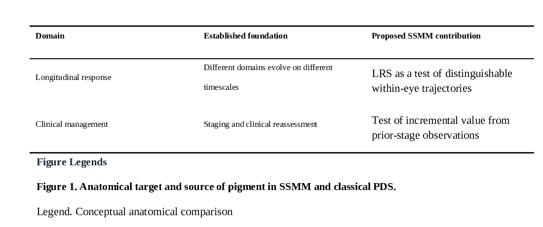
Original tables (Tables 1–4)
These are unmodified table extracts from the original 19-page manuscript. For selectable text and exact page context, open the original PDF at page 15.
Show all four original tables


Original figures
The following diagrams are reproduced by cropping the unchanged preprint PDF; see the original figure legends in the manuscript.