Biological Basis and Limitations of Permanence in Laser Iris Depigmentation
A scientific examination of how changes in anterior iris stromal pigmentation influence visible eye color, why biological pigment reduction and optical stability are distinct outcomes, and how residual pigmentation, stromal architecture, and longitudinal measurement define the limits of permanence.
Quick Answer
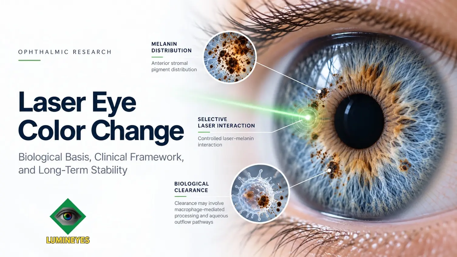
The biological basis of laser iris depigmentation is the intended reduction of optically significant melanin within the anterior iris stroma. By modifying the contribution of superficial pigmentation to light absorption, such treatment may alter the apparent color of the underlying iris. The visible outcome also depends on residual pigment, tissue architecture, and optical scattering, rather than pigment reduction alone.
Permanence is not a single biological endpoint. A lasting reduction in stromal pigmentation, a stable visible iris color, and sustained ocular health represent three related but independently measurable outcomes. Residual pigmentation may remain optically important even after substantial lightening, while an apparent change in color over time does not necessarily establish true melanin regeneration.
Accordingly, long-term results are best interpreted through repeated anatomical and optical observations, with clinical safety assessed separately. Neither a predetermined final color nor lifelong invariability of the visible phenotype can be inferred solely from the initial pigment response.
1. Introduction: Defining Biological Permanence
The concept of permanent iris color modification is frequently presented as though the removal of pigment and the resulting optical appearance were equivalent events. In reality, the relationship is more complex. Iris color is an emergent property of pigmentation, tissue organization, and light interaction, whereas melanin reduction is a biological change occurring within a specific anatomical compartment. The two processes are causally related, but they cannot be measured or interpreted using identical criteria.
Laser iris depigmentation is intended to modify pre-existing pigmentation rather than introduce an artificial colorant. This differentiates its anatomical rationale from intraocular cosmetic implants and corneal pigmentation procedures. Nevertheless, the absence of implantation does not establish a particular degree of tissue selectivity, treatment efficacy, or long-term safety. The available scientific literature requires these questions to be considered independently. [7] [8]
The purpose of this review is to examine the biological meaning of permanence after iris pigment modulation. Particular attention is given to the distinction between residual melanin and new pigment formation, the optical consequences of stromal heterogeneity, the time-dependent interpretation of treatment response, and the measurements required to demonstrate long-term stability. Detailed treatment kinetics and procedural safety algorithms are addressed in separate research frameworks.
2. Anatomical Basis of Iris Pigmentation
The human iris is a multilayered structure composed of an anterior border region, a vascular and connective-tissue stroma, smooth-muscle components, and posterior pigmented epithelial layers. Although these compartments collectively contribute to normal iris physiology, their roles in determining visible color are not equivalent. The distribution and concentration of melanin within the anterior stroma are particularly important because they influence the absorption of incident light before it is returned toward the observer.
Histological observations indicate that differences in normal iris color are not adequately explained by variation in the total number of superficial stromal melanocytes. Instead, the intracellular abundance and distribution of melanosomes are important determinants of pigmentation. Electron microscopy has demonstrated measurable differences in melanosomal content across iris color groups, providing an anatomical basis for the distinction between pigment quantity and melanocyte number. [1] [2]
This distinction has implications for interpreting laser-induced lightening. An alteration in the optical influence of melanin does not necessarily require complete destruction or removal of the cells that originally contained it. Conversely, the persistence of melanocytes does not establish that the original pigment burden, spatial distribution, or optical appearance will necessarily be restored after treatment.
2.1 Structural color and residual absorption
Blue and gray iris appearances arise largely from the interaction of incident light with relatively low-pigment anterior tissues and their microscopic architecture. Melanin attenuates portions of the returning light, while stromal microstructure contributes to wavelength-dependent scattering. The resulting phenotype is therefore more complex than a simple relationship between the absolute amount of brown pigment and the apparent color.
Rayleigh-type scattering is commonly invoked as an explanatory model, but the human iris is neither an optically homogeneous medium nor an ideal Rayleigh-scattering system. Stromal fibers, crypts, variable tissue thickness, local pigment deposits, and heterogeneous cellular structures contribute to the observed optical response. Green and hazel appearances particularly illustrate how pigment distribution and structural characteristics can produce intermediate or spatially heterogeneous phenotypes.
Melanin reduction and melanocyte elimination are not synonymous. The quantity of optically active pigment, its intracellular or extracellular location, and its spatial distribution must be distinguished from the number or viability of pigment-containing cells.
The broader organization of the iris is discussed in the dedicated iris stroma and anatomy resource. Here, the relevant anatomical question is how changes in superficial pigmentation modify the visible optical phenotype without establishing that all deeper pigment or pigment-producing cells have disappeared.
3. From Laser–Melanin Interaction to Biological Response
The physical interaction between laser energy and melanin-containing tissue depends on wavelength, pulse duration, energy distribution, absorption, and the optical properties of the target. Selective photothermolysis provides a foundational description of preferential energy absorption within pigmented structures, although the balance between photothermal, photomechanical, and photoacoustic processes varies among laser systems and exposure conditions. [3]
In laser iris depigmentation, the intended biological target is optically relevant pigment within the anterior iris. The resulting response cannot be reduced to a single instantaneous color-conversion event. Initial laser interaction may be followed by pigment-associated changes, cellular processing, local tissue recovery, and evolving optical appearance. These events occur within interconnected biological systems but may follow different temporal trajectories.
Within the Lumineyes™ framework, selective stromal melanin modulation (SSMM) describes the intended controlled interaction with pre-existing anterior stromal pigment. The term defines a proposed targeting and clinical assessment concept; it does not imply that absolute microscopic selectivity has been independently demonstrated. The detailed conceptual boundaries are addressed in the selective stromal melanin modulation framework.
Physical Interaction
Laser energy interacts with susceptible melanin-bearing structures. The immediate physical effect may alter pigment organization, but it does not independently determine the final visible iris color.
Biological Processing
Pigment-associated material may undergo cellular uptake, redistribution, or transport. Macrophage-mediated processing is a plausible contributory mechanism, although its precise quantitative role after iris depigmentation has not been established.
Optical Stabilization
The visible phenotype may continue to evolve as tissue conditions change and residual pigment exerts its optical effects. Biological response and observable color stabilization need not reach their endpoints simultaneously.
A major consequence of this temporal distinction is that an early clinical photograph cannot establish the eventual extent of pigment modification or the stability of the final phenotype. Changes in color may be associated with biological processing, residual pigment distribution, tissue recovery, or combinations of these factors. Their relative contributions cannot be assigned without appropriate longitudinal measurements.
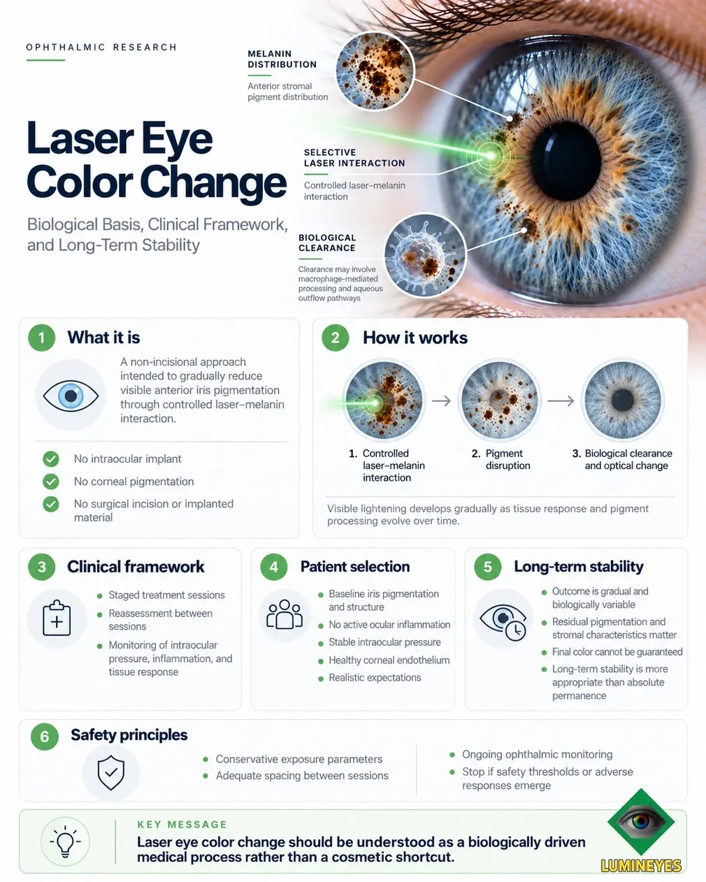
The separate pigment-clearance kinetics research examines pigment release, clearance capacity, and aqueous outflow considerations in greater detail. The present review instead addresses the biological and optical interpretation of a pigment-modification outcome once its initial response has occurred.
4. Biological Determinants of Long-Term Color Stability
A durable alteration in superficial pigmentation may produce a lasting change in iris appearance. However, the magnitude and character of that appearance depend on additional biological variables. The most important distinction is between the amount of pigment modified by treatment and the amount of pigment that remains optically influential afterward.
Residual pigmentation may persist at different depths or in different iris regions. A darker peripupillary zone, sectoral pigment heterogeneity, or comparatively dense deeper stromal pigmentation may continue to affect the visible phenotype even where superficial lightening has occurred. Such patterns do not necessarily indicate regeneration of previously modified pigment. They may instead reflect pigment that was present throughout the observation period.
Stromal architecture introduces another independent source of variability. Tissue thickness, collagen organization, crypt distribution, and the arrangement of the anterior iris can influence the optical result associated with a given pigment burden. Two eyes with apparently similar superficial lightening may consequently exhibit different degrees of brightness, hue, or spatial uniformity. The relative importance of these variables requires prospective structural and optical characterization.
| Determinant | Biological or optical relevance | Implication for permanence |
|---|---|---|
| Residual superficial melanin | Remaining pigment continues to absorb light within the anterior iris. | May limit the degree of visible lightening without representing pigment recurrence. |
| Deeper stromal pigment | Pigment below the principal modified region may remain optically relevant. | Can influence the final hue and apparent depth of coloration. |
| Regional pigment distribution | Peripupillary and sectoral differences produce nonuniform absorption. | May explain persistent heterogeneity despite a durable superficial response. |
| Stromal architecture | Collagen organization, crypts, and tissue thickness influence scattering. | May affect color stability independently of substantial changes in melanin quantity. |
| Biological tissue response | Changes in the local tissue environment may modify the observed phenotype. | Can contribute to optical evolution during the post-treatment interval. |
| Measurement conditions | Lighting, pupil size, exposure, and viewing geometry modify apparent color. | May create apparent differences without demonstrating biological repigmentation. |
These determinants show why an exact final color cannot be calculated solely from the extent of visible pigment reduction. Although stromal pigmentation is a major component of iris coloration, the final appearance is produced by the interaction of multiple anatomical and optical factors. A biologically persistent pigment change may therefore coexist with a phenotype whose measured appearance varies according to regional tissue properties or observation conditions.
5. Limitations of Permanence: Residual Pigment, Repigmentation, and Optical Drift
The concept of permanence becomes clinically meaningful only when the underlying endpoint is specified. A reduction in the amount of visible pigment may persist for an extended period, but this does not establish that the same apparent iris color will remain unchanged throughout life. Nor does a later difference in appearance necessarily demonstrate new melanin synthesis or the return of pigment that had previously been removed.
5.1 Residual pigmentation is not repigmentation
Residual pigmentation refers to melanin that remains within optically relevant iris tissue after an intervention. This material may be superficial, regionally heterogeneous, or located deeper within the stroma. Its continued optical influence can become more noticeable as the surrounding phenotype changes or as regional contrast increases.
True repigmentation, by contrast, would require evidence of a subsequent increase in melanin burden relative to an appropriately documented post-treatment state. Demonstrating such a process would require serial measurements capable of distinguishing pigment accumulation from changes in illumination, tissue appearance, or the visibility of pre-existing pigment.
5.2 The continuing biological capacity of melanocytes
The adult iris ordinarily maintains a relatively stable pigmentation pattern. Nevertheless, its pigment-producing cells remain part of living tissue, and pigmentation may change under certain pathological or pharmacological conditions. Published observations of acquired iris pigmentation changes indicate that adult pigment regulation cannot be assumed to be absolutely immutable. [1]
This does not establish that clinically meaningful pigment regeneration routinely occurs after laser iris depigmentation. The survival of melanocytes, their potential capacity for melanin synthesis, the persistence of residual melanosomes, and any theoretical cellular repopulation are separate questions. Their relevance to delayed color changes must be evaluated experimentally rather than inferred from appearance alone.
5.3 Optical drift without proven pigment regeneration
A change in measured or perceived iris hue may occur without a demonstrated increase in pigment mass. Differences in pupil diameter, regional contrast, stromal reflectance, camera settings, and observation geometry may alter the apparent color. Tissue-related changes over time may also modify the relationship between existing pigment and light scattering.
Accordingly, the term optical drift may be useful descriptively for a longitudinal change in the recorded phenotype whose biological cause has not yet been established. It should not be used interchangeably with repigmentation.
A darker appearance after treatment does not automatically demonstrate melanin regeneration. The possibilities include persistent residual pigment, changes in the optical contribution of pre-existing pigment, measurement variability, tissue-related changes, and genuine new pigment accumulation. These explanations require different evidence.
| Observed phenomenon | Possible interpretation | Evidence required |
|---|---|---|
| Persistent darker iris sectors | Regional residual pigmentation or heterogeneous stromal optics. | Baseline and longitudinal sectoral imaging under comparable conditions. |
| Delayed lightening | Continuing biological response or evolution of tissue optical properties. | Serial pigment-sensitive and colorimetric observations. |
| Apparent darkening | Measurement variation, altered optical contrast, or possible pigment increase. | Reproducible imaging and, where possible, independent pigment assessment. |
| Documented pigment increase | Potential new pigment accumulation or redistribution. | Objective longitudinal evidence distinguishing the location and origin of pigment. |
The distinction between these patterns is essential to interpreting long-term clinical observations. Without a documented post-treatment reference state and comparable follow-up measurements, the biological cause of a later color difference may remain uncertain even when that difference is visually apparent.
6. Three Independent Endpoints of Long-Term Stability
For clinical research purposes, permanence should not be regarded as a binary designation. It is more informative to distinguish the persistence of pigment modification from the stability of the optical phenotype and from longitudinal ocular health. These dimensions may evolve differently and require different measurement strategies.
Biological Pigment Stability
The persistence of a measurable reduction in optically relevant iris pigmentation relative to a defined baseline or post-treatment reference.
Optical Phenotype Stability
The reproducibility of iris appearance across comparable observation conditions, including regional color distribution, brightness, and hue.
Ocular Clinical Stability
The longitudinal status of intraocular pressure, inflammation, iris anatomy, endothelial parameters, and relevant visual function.
A stable visible phenotype can provide evidence of optical durability, but it cannot independently establish the precise histological fate of modified pigment. Similarly, a documented pigment decrease does not establish that the measured color will remain identical under all future conditions. Finally, neither of these observations is a substitute for clinical monitoring of anterior-segment health.
Long-term optical stability may be defined as the persistence of a reproducible iris color phenotype over serial observations obtained using standardized imaging conditions, with biological pigment stability and ocular safety assessed as separate endpoints.
This operational approach is more useful than an unsupported claim of absolute lifelong permanence. It allows different aspects of an outcome to be compared, quantified, and investigated while preserving the distinction between a visible result and its biological basis.
7. Measuring Permanence: From Photographs to Reproducible Evidence
Demonstrating long-term optical stability requires more than a pair of photographs taken before and after treatment. A scientifically meaningful comparison must account for acquisition conditions, anatomical configuration, regional pigment heterogeneity, and the interval between observations. Without these controls, apparent changes may reflect imaging variability rather than a genuine biological transition.
7.1 Standardized iris color assessment
Imaging protocols should seek to control illumination spectrum and intensity, camera calibration, exposure, white balance, viewing geometry, and pupil diameter. Photographic conditions should be reported consistently, and color measurements should be evaluated using reproducible methods. Where appropriate, calibrated colorimetric coordinates and regional measurements may provide a more informative description than subjective shade labels alone.
Uniform whole-iris averages may obscure clinically relevant regional differences. For this reason, central, intermediate, and peripheral iris regions may require separate evaluation, particularly in eyes with peripupillary pigment concentration, sectoral heterogeneity, or asymmetric lightening. Such measurements should also distinguish the iris from reflections, eyelid shadows, and other photographic artifacts.
7.2 Structural and biological assessment
Clinical imaging may support evaluation of iris configuration, tissue thickness, and other anterior-segment characteristics. However, a structural image or visible color measurement is not necessarily a direct quantitative assay of melanin. The capacity of any imaging method to estimate pigment burden must be established through appropriate validation.
A robust longitudinal framework should therefore distinguish what each technique actually measures. Photography and colorimetry primarily describe the optical phenotype; anterior-segment imaging describes aspects of structure; and clinical examination identifies safety findings. Correlations among these domains are scientifically valuable, but one should not automatically be treated as a substitute for another.
| Measurement domain | Proposed observation | Principal interpretive limit |
|---|---|---|
| Colorimetry | Calibrated overall and regional color measurements. | Does not directly identify the cellular source of pigment. |
| Serial photography | Standardized records of visible iris phenotype over time. | Sensitive to lighting, pupil size, and camera variation. |
| Anterior-segment imaging | Iris geometry, thickness, and selected tissue characteristics. | Structural signal is not automatically equivalent to melanin concentration. |
| Clinical examination | IOP, inflammation, pupil function, and other relevant findings. | Does not independently quantify optical permanence. |
| Longitudinal comparison | Repeated observations using predefined endpoints. | Requires complete follow-up and consistent acquisition methods. |
A future prospective study could use predefined measurement intervals and quantitative change thresholds to distinguish an early response phase from a relatively stable optical phase. The thresholds themselves would require methodological validation; they should not be assumed to represent a universal biological definition of permanence.
This distinction provides a useful bridge between descriptive clinical photography and quantitative ophthalmic research. The objective is not merely to document that an iris looks different, but to determine whether the difference is reproducible, regionally consistent, biologically interpretable, and sustained under comparable observation conditions.
8. Clinical Boundaries: Stability Does Not Establish Safety
The scientific interpretation of pigment permanence must remain separate from the assessment of ocular safety. Laser exposure directed at iris pigmentation may be associated with pigment release, transient or persistent inflammatory findings, intraocular pressure changes, and other anterior-segment responses. The absence of a surgical incision does not remove the need to evaluate these physiological events.
Pigment dispersion syndrome and pigmentary glaucoma provide established examples of how pigment-associated trabecular dysfunction can affect aqueous outflow. Although naturally occurring pigment dispersion cannot be regarded as directly equivalent to laser-induced pigment release, its physiology supports the importance of monitoring pressure and outflow-related findings after iris-directed intervention. [4]
Peer-reviewed case literature has reported severe secondary pigmentary glaucoma after cosmetic iris laser treatment. Such reports establish the possibility of serious adverse outcomes but do not determine their incidence across heterogeneous laser systems or clinical protocols. [6]
Corneal endothelial assessment also remains relevant because endothelial reserve and cellular morphology are clinically important components of anterior-segment health. Specular microscopy provides measurements that can support longitudinal endothelial evaluation, although the interpretation of serial cell counts requires attention to measurement variability and image quality. [5]
Optical permanence and ocular safety are independent clinical questions. A stable iris color does not demonstrate normal trabecular function, endothelial stability, or the absence of persistent inflammation. These outcomes require their own defined observations.
The retrospective observations associated with the MyLumineyes clinical framework are documented separately in the longitudinal clinical data record. Those observations should be interpreted according to their study design, follow-up completeness, and predefined outcome measures rather than extrapolated into universal safety estimates.
9. Evidence Boundaries and Future Research Priorities
The anatomical foundations of human iris pigmentation are supported by histological and morphological studies, while selective laser absorption is grounded in established laser–tissue biophysics. These bodies of evidence provide a scientific basis for examining iris pigment modulation. They do not, however, independently establish the frequency, magnitude, or lifelong durability of clinical outcomes after elective laser iris depigmentation. [1] [3]
Contemporary reviews of cosmetic eye color modification identify important differences among the available interventions and continue to highlight limitations in the comparative and longitudinal evidence for laser-based techniques. The evidence is insufficient to assign a universal long-term permanence rate or complication rate to the heterogeneous category of laser iris depigmentation. [7] [8]
Future work should address not merely whether a visible change persists, but which biological and optical mechanisms explain its persistence. This will require consistent baseline characterization, repeatable measurements, adequate follow-up intervals, and explicit separation of direct observations from mechanistic hypotheses.
Residual Pigment Mapping
Determine whether anatomical and optical measurements can characterize residual stromal pigment distribution reliably after treatment.
Repigmentation Assessment
Develop longitudinal criteria capable of distinguishing genuine new pigment accumulation from residual pigmentation, redistribution, and optical variability.
Optical Stability Metrics
Establish reproducible colorimetric and regional imaging endpoints for quantifying phenotype stability over extended follow-up.
Clinical Correlation
Examine associations between pigment response, anatomical characteristics, intraocular pressure, inflammation, endothelial measurements, and long-term optical outcomes.
These priorities also require a clear hierarchy of evidence. Case reports may identify clinically important possibilities, retrospective cohorts may characterize observed patterns, and prospective longitudinal studies may evaluate predefined associations. None of these designs should be interpreted beyond the scope of the observations they can support.
The methodological principles underlying such distinctions are discussed in How Clinical Evidence Is Evaluated. The present review provides the biological and optical rationale for defining the endpoints that future studies should measure.
10. Conclusion: The Meaning and Limits of Biological Permanence
The scientific basis of laser iris depigmentation lies in the relationship between melanin-containing anterior stromal tissue and the optical mechanisms responsible for visible iris color. Modifying superficial pigmentation can change the balance between light absorption and scattering, but the resulting appearance remains dependent on the distribution of residual melanin and the architecture of the underlying tissue.
Biological permanence should therefore not be equated with absolute optical immutability. A persistent reduction in pigment burden may support a durable change in iris appearance, while the final phenotype may still be influenced by residual pigmentation, regional tissue properties, and longitudinal biological or optical variability. A subsequent darker appearance cannot, without objective evidence, be classified automatically as melanin regeneration.
The most useful long-term framework separates three endpoints: persistent pigment modification, reproducible optical phenotype, and sustained ocular clinical stability. Their integration through standardized imaging, structural assessment, and longitudinal ophthalmic monitoring offers a more rigorous basis for investigating permanence than a binary distinction between temporary and permanent color change.
Ultimately, the limits of permanence are best defined not by an absolute promise concerning the iris phenotype, but by the biological mechanisms that can be documented, the optical changes that can be measured, and the clinical outcomes that remain stable during follow-up.
References and Scientific Literature
- Imesch PD, Wallow IH, Albert DM. The color of the human eye: a review of morphologic correlates and of some conditions that affect iridial pigmentation. Survey of Ophthalmology. 1997;41(Suppl 2):S117–S123. DOI: 10.1016/S0039-6257(97)80018-5
- Imesch PD, Bindley CD, Khademian Z, et al. Melanocytes and iris color: electron microscopic findings. Archives of Ophthalmology. 1996;114(4):443–447. DOI: 10.1001/archopht.1996.01100130439015
- Anderson RR, Parrish JA. Selective photothermolysis: precise microsurgery by selective absorption of pulsed radiation. Science. 1983;220(4596):524–527. DOI: 10.1126/science.6836297
- Scuderi G, Contestabile MT, Scuderi L, et al. Pigment dispersion syndrome and pigmentary glaucoma: a review and update. International Ophthalmology. 2019;39(7):1651–1662. DOI: 10.1007/s10792-018-0938-7
- Chaurasia S, Vanathi M. Specular microscopy in clinical practice. Indian Journal of Ophthalmology. 2021;69(3):517–524. DOI: 10.4103/ijo.IJO_574_20
- Ong AY, Ching-A-Sue G, Krilis M, et al. Refractory iatrogenic pigmentary glaucoma secondary to cosmetic laser treatment: a case report. European Journal of Ophthalmology. 2023;33(2):NP1–NP4. DOI: 10.1177/11206721211050338
- D'Oria F, Abu-Mustafa SK, Alio JL. Cosmetic change of the apparent color of the eye: a review on surgical alternatives, outcomes and complications. Ophthalmology and Therapy. 2022;11:465–477. DOI: 10.1007/s40123-022-00458-2
- D'Oria F, Alio JL. Surgical techniques for cosmetic eye color change: a narrative review. Ophthalmology and Therapy. 2025;14:1685–1694. DOI: 10.1007/s40123-025-01177-0
