Full manuscript · Chapter 1 of 6
Selective Stromal Melanin Modulation (SSMM)
Associated research DOI: https://doi.org/10.5281/zenodo.24203344. Exact manuscript–record relationship awaits verification.
Selective Stromal Melanin Modulation (SSMM): an integrative biophysical and clinical framework for response-guided laser iris pigment modification
Mustafa Mete, MD
MyLumineyes Research, Istanbul, Türkiye
Corresponding author: Mustafa Mete, MD — info@mylumineyes.com
ORCID: 0009-0004-3128-2853
Abstract
Purpose: To synthesize the optical, anatomical and longitudinal concepts underlying selective stromal melanin modulation (SSMM) and formulate experimentally testable questions for laser-directed iris pigment modification.
Methods: A critical narrative synthesis of iris biology, optical tissue interaction, published laser-related evidence and author-reported longitudinal clinical observations was used to develop a conceptual Target–Response–Decision architecture. Evidence from published studies was distinguished from proposed mechanisms and clinical-program estimates, which were not analyzed as an independently audited cohort.
Results: The SSMM framework separates biophysical, anatomical and functional selectivity; initial pigment modification, subsequent pigment processing and tissue response; and observations obtained at different treatment stages. Longitudinal Response Selectivity (LRS) describes the proposed distinguishability of intended pigment changes from accompanying ocular response trajectories. Seven falsifiable propositions address targeting, structural effects, baseline phenotype, response timing, staged decisions and physiological outcomes. No controlled evidence of selective targeting, validated prediction model or quantified treatment efficacy is supplied by this conceptual synthesis.
Conclusion: SSMM integrates established principles and clinical hypotheses into an explicit research architecture. Its proposed selectivity and response-guided value require independent optical, biological and longitudinal clinical evaluation.
Keywords: iris stroma; selective stromal melanin modulation; laser–tissue interaction; longitudinal response selectivity; iris pigmentation; anterior-segment imaging
Introduction
Human iris coloration reflects melanin content, pigment distribution, stromal architecture, optical absorption, and light scattering. Morphological investigations demonstrate that visible variation in normally pigmented irides is related substantially to melanin content and distribution rather than melanocyte number alone [1, 2].
The anterior iris stroma contains melanocytes, fibroblasts, vascular and neural structures, and extracellular matrix components. Pigment-directed intervention therefore takes place in tissue with anatomical and physiological roles extending beyond coloration [3, 4].
Selective photothermolysis provides a physical foundation for preferential interaction with an absorbing chromophore [5]. Tissue optics emphasizes the importance of wavelength, local absorption, scattering, and tissue structure [6]. Depending on exposure conditions, laser interaction may involve photothermal and photomechanical effects [7].
Experimental laser exposure has produced measurable changes in iris pigmentation and associated cellular responses in a rabbit model [8]. Human clinical work has described staged laser iris depigmentation with repeated assessment and imaging [9]. Broader ophthalmic reviews place these and other cosmetic eye-color interventions within a heterogeneous evidence base [10, 11]. Sharing a visible endpoint does not establish equivalence among their mechanisms or exposure architectures.
Laser iris depigmentation names a visible endpoint, without specifying the optical target, pigment processing, tissue responses, or longitudinal clinical decisions. SSMM was developed within Lumineyes research to integrate these domains [12].
Scientific background and narrative synthesis
Lumineyes developed from exploratory laser-directed iris pigment modification beginning in 2009, with later technical and clinical refinement. The 2017–2026 program enabled repeated assessment across treatment stages.
SSMM addresses four questions: how baseline pigmentation affects response; how optical interaction relates to pigment change; how intended pigment change differs from accompanying tissue responses; and whether earlier-stage observations inform subsequent decisions.
This conceptual and mechanistic synthesis integrates iris biology, laser–tissue science, ophthalmic literature, and longitudinal clinical experience. Clinical observations motivate hypotheses rather than representing a newly audited cohort.
Operational Definition and Three Dimensions of Selectivity
Selective stromal melanin modulation is an intended strategy of preferential laser interaction with optically relevant melanin-bearing structures of the anterior iris stroma, directed toward progressive pigmentary modification through clinically monitored, staged intervention, without deliberate ablation of supporting stromal architecture.
Selective means preferential, not exclusive, interaction; stromal specifies the target compartment; modulation distinguishes pigment change from extensive stromal ablation.
Three dimensions are defined: biophysical selectivity concerns chromophore-directed optical interaction; anatomical selectivity concerns distribution and persistence of structural effects; functional selectivity concerns ocular physiology. Each requires independent evidence beyond visible color change.
Biophysical Foundations of Stromal Pigment Modification
Melanin and the local optical environment
Melanin is an endogenous ocular chromophore whose interaction with optical radiation depends on absorption, wavelength, exposure conditions, and the surrounding tissue environment [4–6]. Within SSMM, melanin-bearing structures of the anterior iris stroma constitute the intended target.
Pigment distribution, architecture, scattering, and spatial exposure characteristics modify response; similarly colored eyes may therefore have different local optics and pigmentary outcomes.
Localized pigment interaction
Pigmented-tissue exposure may produce photothermal and photomechanical effects [7], potentially including microcavitation. Its occurrence and stromal consequences under SSMM-specific conditions require direct characterization.
Thermal and stress confinement depend on pulse duration relative to heat-diffusion and acoustic-transit times. Melanosome and retinal experiments show that pulse shape alters microcavitation thresholds and bubble dynamics [13]; such thresholds are not validated limits for human iris exposure. SSMM-specific pulse geometry, pigment distribution, and tissue effects require direct measurement.
Direct experimental iris evidence
Yildirim and colleagues studied 532-nm Q-switched Nd:YAG exposure in 12 pigmented rabbits and reported reduced anterior stromal pigmentation accompanied by increased stromal pigment-laden macrophages [8]. Histological changes in the posterior pigment epithelium were not identified under those experimental conditions. The findings demonstrate a distinction between anterior stromal pigment alteration and subsequent cellular processing; they do not establish equivalence with the Lumineyes clinical exposure protocol.
Controlled Pigment Response: Modification, Processing, and Recovery
Controlled anterior stromal pigment release denotes planned targeting, staged clinical assessment, and revised decisions, not directly quantified liberated melanin mass.
Three distinct processes are involved:
Pigment modification follows initial melanin interaction; pigment processing includes liberation, redistribution, cellular handling, and possible clearance; tissue recovery comprises subsequent cellular, matrix, anatomical, and functional changes. Their trajectories may overlap or diverge.
Visible lightening does not quantify pigment clearance or prove restoration of melanocyte function and extracellular matrix. P-CLEAR addresses release and processing [14]; cellular and matrix responses remain separate questions.
Human iris biology supports these distinctions: Smith-Thomas et al. found fibroblast-dependent pigmentary responses of iridial melanocytes to prostaglandin analogues [15], while Lan et al. identified metalloproteinases and inhibitors in human iris and ciliary body [16]. Both findings support investigating cellular interaction and matrix regulation but do not directly establish analogous post-laser responses.
Distinction From Classical Pigment Dispersion Syndrome
Classical pigment dispersion syndrome (PDS) principally involves pigment liberation from the posterior iris pigment epithelium, commonly in association with mechanical iris–zonular interaction [17]. SSMM instead concerns deliberate laser interaction with melanin-bearing structures of the anterior iris stroma. The anatomical distinction is illustrated in Figure 1.
The initiating source and mechanism differ, although downstream pigment-processing pathways may overlap. Their distinction does not imply complete biological separation.
Serious complications, including refractory pigmentary glaucoma and paracentral acute middle maculopathy, have been reported following laser procedures for cosmetic iris color change [18]. Such case reports establish clinically relevant events but do not quantify their frequency or demonstrate equivalence across different exposure protocols. This distinction strengthens the rationale for protocol-specific and longitudinal outcome assessment.
The Lumineyes™ Conceptual Fingerprint
The Lumineyes Conceptual Fingerprint connects Target → Response → Decision: intended pigment-bearing anatomy and optical targeting; ensuing pigment and biological responses; and use of longitudinal observations for subsequent stages.
Staged intervention and reassessment predate SSMM [9]. Its contribution is integrating them with three selectivity dimensions, controlled pigment response, and LRS.
Selective laser trabeculoplasty offers a mechanistic parallel with a different target: Alvarado et al. observed monocyte recruitment into the trabecular meshwork after SLT, increased outflow facility after macrophage infusion, and higher Schlemm canal endothelial conductivity after monocyte-associated factors [19]. These observations justify study of potentially convergent downstream responses after SSMM, without establishing that the same pathway operates.
Phenotype and Interindividual Response Variability
Visible color cannot quantify regional melanin distribution or optical accessibility; similar colors may conceal distinct stromal architecture.
The Lumineyes Iris Pigmentation Scale (LIPS) describes apparent anterior stromal pigmentation through a proposed five-grade architecture [20]. Grades I–IV represent increasing diffuse apparent pigmentation; Grade V identifies substantial spatial heterogeneity and is not a more densely pigmented grade than Grade IV. This distinction should not be conflated with different grading schemes in which the final grade denotes the darkest pigmentation [9].
Lumineyes Optical Architectural Typology (L-OAT) explores optical and architectural phenotype [21]. Whether baseline features predict response remains to be validated.
Longitudinal Response Selectivity (LRS)
Definition and position
Longitudinal Response Selectivity (LRS) — Proposed within the SSMM framework (2026).
Longitudinal Response Selectivity (LRS) is a proposed concept within the SSMM framework describing the extent to which intended stromal pigment modification can be distinguished from accompanying tissue responses across successive observation periods and treatment stages.
LRS extends biophysical, anatomical, and functional assessment through repeated observations (Figure 2). It describes longitudinal differentiation of pigment and tissue trajectories, not a fourth optical mechanism or proof of tissue preservation.
Temporal interpretation
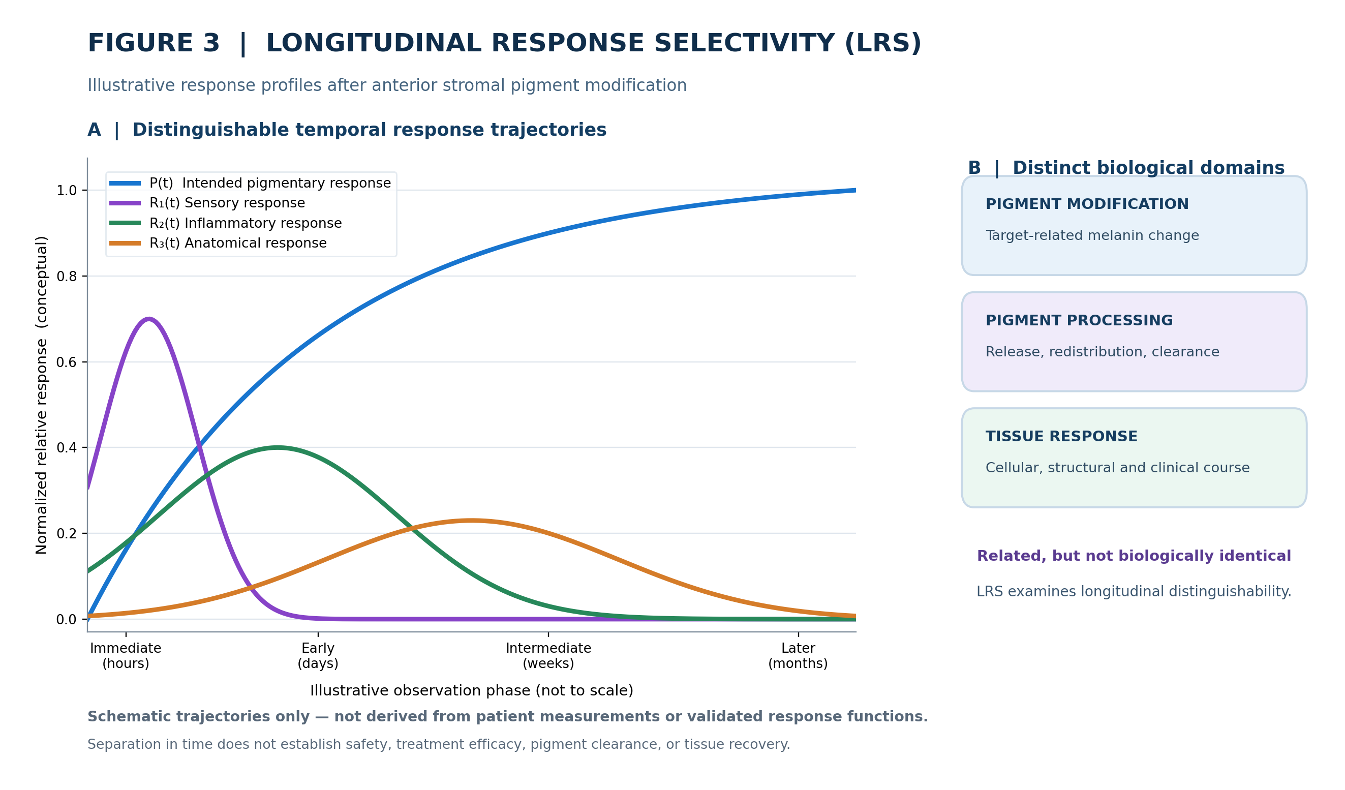
Laser interaction is brief, whereas pigmentary, sensory, anatomical, and inflammatory trajectories may evolve separately. A single examination can miss these differences (Figure 3).
Temporal Response Architecture (TRA) organizes response domains across time [22]. LRS asks whether pigmentary and tissue trajectories can be distinguished; aqueous cells and flare are candidate inflammatory measures, not established SSMM-specific kinetics.
Operational assessment
Let P(t) denote observed intended pigment change at time t and R₁(t), R₂(t), …, Rₙ(t) denote accompanying response domains. LRS asks whether trajectories differ in onset, magnitude, duration, persistence, or stage dependence.
LRS requires baseline data, paired repeated observations, stage timestamps, appropriate outcomes, and models of within-eye and within-patient correlation. Exposure and medication histories must contextualize trajectories.
No numerical LRS index is proposed; separable trajectories do not alone establish favorable biological outcomes.
Longitudinal clinical observations motivating SSMM hypotheses
Program scale and follow-up
The contemporary Lumineyes program, principally spanning 2017–2026, comprises approximately 1,500 patients and 3,000 distinct treated eyes according to the author's clinical-program estimates. Repeated treatment sessions in the same eye are not counted as additional treated eyes.
Based on clinical-program estimates, approximately 100 patients have follow-up exceeding five years, and at least 10 have reached or approached nine years of observation. These are approximations from the clinical archive, not endpoints derived from a completed patient-level outcomes analysis with verified denominators or uniform follow-up schedules.
Evolution of staged management
The method underwent progressive refinement in patient selection, treatment organization, postoperative management, and repeated reassessment. In the author's experience, later phases were associated with fewer clinically observed transient reactions, apart from transient photophobia. These observations contributed to the response-guided clinical architecture.
Transient photophobia-associated discomfort
Light sensitivity was frequently reported during staged treatment. Direct questioning of hundreds of patients suggested that discomfort sometimes described as pain was predominantly associated with light exposure rather than spontaneous ocular pain. Photophobia typically emerged after the second or third session and was relatively uncommon after the first. Affected patients often preferred darkness or kept their eyes closed for approximately one to two hours, after which symptoms generally resolved.
Transient corneal findings
Subtle Descemet membrane folds were occasionally observed, particularly in older patients and during treatment courses associated with substantial pigment liberation. These findings could remain detectable over a seven-to-ten-day course. Among affected patients examined approximately four months later, residual folds and persistent edema were not observed. The precise time of resolution between examinations was not systematically established.
Inflammatory observations
Mild iridocyclitis and fibrinous responses were recalled predominantly from earlier phases of clinical development. Some episodes appeared to coincide with incomplete adherence to prescribed topical corticosteroid treatment; fewer inflammatory events were recalled among selected patients receiving additional systemic support. These associations were not established through controlled comparisons. Reported episodes resolved with clinical treatment during available follow-up.
Other longitudinal observations
Approximately 15 patients were recalled to have experienced early postoperative intraocular-pressure elevation, reportedly resolving with medical treatment. The author recalls no persistent clinically apparent sequelae among affected patients returning for examination during available follow-up, although observation duration and examination completeness varied. Quantitative corneal endothelial assessment remains a priority, especially in older individuals and those with substantial cumulative exposure.
Contribution and Complementary Research Frameworks
SSMM combines established optical and biological principles into a testable anterior stromal model linking target specificity, three selectivity domains, pigment processing, longitudinal differentiation, and adaptive decisions.
Complementary proposed frameworks address phenotype (LIPS, L-OAT), pigment processing (P-CLEAR), temporal responses (TRA), and pre-exposure safety states (L-SAFE) [25]. Stromal biology, blood–aqueous barrier dynamics, and phenotype–response coupling remain research priorities; L-TIDE separately addresses multimodal mechanistic identifiability.
Independent optical, biological, and clinical evidence—not cross-citation among proposals—must test SSMM.
Testable Propositions
Proposition 1 — Target interaction. Under defined experimental conditions, anterior stromal melanin-bearing structures exhibit a distinguishable optical response relative to suitable surrounding tissue components.
Proposition 2 — Pigmentary versus anatomical response. The magnitude and distribution of visible pigmentary change need not correspond directly to accompanying stromal structural alteration.
Proposition 3 — Phenotype dependence. Baseline pigmentation pattern and optical architecture may explain variation in subsequent pigmentary outcomes beyond broad color labels.
Proposition 4 — Longitudinal differentiation. Intended pigmentary and accompanying tissue responses can be assessed for distinguishable onset, magnitude, and recovery trajectories under reliable measurement conditions.
Proposition 5 — Added value of prior-stage observations. Previous-stage observations may contribute predictive information beyond baseline phenotype and cumulative exposure.
Proposition 6 — Long-term tissue response. Anatomical and physiological measurements may vary with age, baseline characteristics, and cumulative exposure.
Proposition 7 — Mechanistic discrimination. Independent domain-specific measurements may improve differentiation among competing explanations for the observed pigmentary change.
These propositions can be evaluated with controlled tissue studies, standardized imaging, appropriate comparators, and repeated clinical measurements. Failure of preferential targeting, reliable longitudinal differentiation, or added predictive information would challenge the corresponding SSMM propositions.
Research Priorities
Quantitative phenotyping. Standardized photography and regional optical measurements can characterize iris pigmentation over time [26]. Baseline classification should be assessed for observer reliability before use as a predictive instrument.
Experimental interaction studies. Defined exposure conditions should be related to pigment change and independently measured stromal responses, including cellular and matrix findings where feasible.
Longitudinal clinical analysis. Patient-level, eye-level, session-level, and event-level observations should be separated. Analyses should account for treatment stages nested within eyes and eyes nested within patients.
Corneal endothelial assessment. Serial endothelial cell density and morphology can provide quantitative information beyond clinical impressions of corneal clarity, particularly after substantial cumulative exposure.
Adaptive assessment. A direct test of the Lumineyes Conceptual Fingerprint is whether information from earlier stages improves subsequent outcome characterization compared with baseline and exposure variables alone.
Discussion
Experimental iris research links pigment alteration with cellular processing [8], while prior clinical work describes staged depigmentation and reassessment [9]. SSMM adds explicit selectivity domains and LRS to connect target, response, and decisions beyond visible lightening.
Unlike classical PDS, SSMM begins at a different anatomical source and mechanism [17]. Fibroblast-dependent melanocyte behavior and matrix regulation support separate evaluation of pigmentary and architectural changes [15, 16], without proving post-laser equivalence.
LRS requires domain-specific measurements: flare photometry quantifies aqueous protein scattering [23], and swept-source AS-OCT shows preliminary associations between particle characteristics and clinically identified pigment or inflammatory cells [24]. Neither substitutes for pigment imaging or endothelial assessment.
Conclusion
Selective stromal melanin modulation defines an integrated framework for studying laser-directed anterior iris stromal pigment modification. Biophysical, anatomical, and functional selectivity provide distinct investigative dimensions, while Longitudinal Response Selectivity addresses the temporal distinguishability of intended pigmentary modification from accompanying tissue responses.
The Selective Targeting → Controlled Pigment Response → Longitudinal Response Selectivity → Adaptive Decision architecture connects the intended optical interaction with subsequent pigment processing, tissue response, and staged clinical management. It translates the clinical development of Lumineyes into explicit research propositions that can be tested independently.
References
[1] Imesch PD, Wallow IHL, Albert DM. The color of the human eye: a review of morphologic correlates and of some conditions that affect iridial pigmentation. Surv Ophthalmol. 1997;41(Suppl 2):S117–S123. https://doi.org/10.1016/S0039-6257(97)80018-5
[2] Wilkerson CL, Syed NA, Fisher MR, Robinson NL, Wallow IHL, Albert DM. Melanocytes and iris color: light microscopic findings. Arch Ophthalmol. 1996;114(4):437–442. https://doi.org/10.1001/archopht.1996.01100130433014
[3] Borrás T. The cellular and molecular biology of the iris, an overlooked tissue. J Glaucoma. 2014;23(8 Suppl 1):S39–S42. https://doi.org/10.1097/IJG.0000000000000104
[4] Hu DN, Simon JD, Sarna T. Role of ocular melanin in ophthalmic physiology and pathology. Photochem Photobiol. 2008;84(3):639–644. https://doi.org/10.1111/j.1751-1097.2008.00316.x
[5] Anderson RR, Parrish JA. Selective photothermolysis: precise microsurgery by selective absorption of pulsed radiation. Science. 1983;220(4596):524–527. https://doi.org/10.1126/science.6836297
[6] Jacques SL. Optical properties of biological tissues: a review. Phys Med Biol. 2013;58(11):R37–R61. https://doi.org/10.1088/0031-9155/58/11/R37
[7] Jacques SL. Laser-tissue interactions: photochemical, photothermal, and photomechanical. Surg Clin North Am. 1992;72(3):531–558. https://doi.org/10.1016/S0039-6109(16)45731-2
[8] Yildirim Y, Duzgun E, Kar T, et al. Evaluation of color-changing effect and complications after Nd:YAG laser application on iris surface. Med Sci Monit. 2016;22:107–114. https://doi.org/10.12659/MSM.895086
[9] Grimaldos Ruiz P. Photoablative cosmetic iridoplasty: effective, safe, and predictable—eye color change in 1176 eyes. Int Ophthalmol. 2021;41:1381–1393. https://doi.org/10.1007/s10792-021-01693-5
[10] D’Oria F, Abu-Mustafa SK, Alio JL. Cosmetic change of the apparent color of the eye: a review on surgical alternatives, outcomes and complications. Ophthalmol Ther. 2022;11:465–477. https://doi.org/10.1007/s40123-022-00458-2
[11] D’Oria F, Alio JL. Surgical techniques for cosmetic eye color change: a narrative review. Ophthalmol Ther. 2025;14:1685–1694. https://doi.org/10.1007/s40123-025-01177-0
[12] Mete M. Selective Stromal Melanin Modulation (SSMM) [preprint]. Zenodo. 2026. https://doi.org/10.5281/zenodo.23424601
[13] Deladurantaye P, Méthot S, Mermut O, Galarneau P, Rochette PJ. Potential of sub-microsecond laser pulse shaping for controlling microcavitation in selective retinal therapies. Biomed Opt Express. 2020;11(1):109–132. https://doi.org/10.1364/BOE.11.000109
[14] Mete M. Pigment Release-Clearance Balance in Selective Stromal Melanin Modulation: A Mechanistic Review and Proposed P-CLEAR Framework [preprint]. Zenodo. 2026. https://doi.org/10.5281/zenodo.23221292
[15] Smith-Thomas L, Moustafa M, Spada CS, et al. Latanoprost-induced pigmentation in human iridial melanocytes is fibroblast dependent. Exp Eye Res. 2004;78(5):973–985. https://doi.org/10.1016/j.exer.2003.12.003
[16] Lan J, Kumar RK, Di Girolamo N, McCluskey P, Wakefield D. Expression and distribution of matrix metalloproteinases and their inhibitors in the human iris and ciliary body. Br J Ophthalmol. 2003;87(2):208–211. https://doi.org/10.1136/bjo.87.2.208
[17] Zeppieri M. Pigment dispersion syndrome: a brief overview. J Clin Transl Res. 2022;8(5):344–350. https://doi.org/10.18053/jctres.08.202205.004
[18] Liu J, Korban S, Moster MR, Rhéaume M-A, Wang Q. Bilateral severe iatrogenic pigmentary glaucoma following laser treatment for cosmetic iris color change. Am J Ophthalmol Case Rep. 2023;32:101927. https://doi.org/10.1016/j.ajoc.2023.101927
[19] Alvarado JA, Katz LJ, Trivedi S, Shifera AS. Monocyte modulation of aqueous outflow and recruitment to the trabecular meshwork following selective laser trabeculoplasty. Arch Ophthalmol. 2010;128(6):731–737. https://doi.org/10.1001/archophthalmol.2010.85
[20] Mete M. The Lumineyes Iris Pigmentation Scale (LIPS): A Proposed Clinical Framework for Grading Anterior Stromal Iris Pigmentation. Zenodo research record. 2026. https://doi.org/10.5281/zenodo.23108463
[21] Mete M. Lumineyes Optical Architectural Typology (L-OAT): A Multimodal Hypothesis for Quantitative Iris Phenotyping. Zenodo preprint. 2026. https://doi.org/10.5281/zenodo.23066772
[22] Mete M. Temporal Response Architecture After Laser-Based Iris Pigment Modification: A Mechanistic Synthesis of Anterior-Segment Response Domains. Zenodo preprint. 2026. https://doi.org/10.5281/zenodo.23128049
[23] Tugal-Tutkun I, Herbort CP. Laser flare photometry: a noninvasive, objective, and quantitative method to measure intraocular inflammation. Int Ophthalmol. 2010;30(5):453–464. https://doi.org/10.1007/s10792-009-9310-2
[24] Bellchambers A, Shu R, Chu CJ, Petrushkin H, Solebo AL. Differentiation of anterior chamber pigment and inflammatory cells using swept-source optical coherence tomography: a cross-sectional study. Eye (Lond). 2025;39:1744–1750. https://doi.org/10.1038/s41433-025-03697-2
[25] Mete M. Lumineyes Anterior-Segment Safety Staging (L-SAFE): A Proposed Framework for Pre-Exposure Assessment in Laser Iris Pigment Reduction. Zenodo research record. 2026. https://doi.org/10.5281/zenodo.23046455
[26] Takamoto T, Schwartz B, Cantor LB, Hoop JS, Steffens T. Measurement of iris color using computerized image analysis. Curr Eye Res. 2001;22(6):412–419. https://doi.org/10.1076/ceyr.22.6.412.5490
Table 1. Three dimensions of selectivity within SSMM
| Dimension | Scientific question | Measurement domain |
| Biophysical | Is interaction preferentially directed toward the intended pigment-bearing target? | Optical and laser–tissue measurements |
| Anatomical | Where do structural effects occur, and how do they evolve? | Structural imaging and anatomical assessment |
| Functional | How do relevant ocular functions behave following exposure? | Clinical and physiological assessment |
Table 2. Candidate longitudinal measurements for evaluating LRS
| Response domain | Candidate observation/measurement | What it establishes |
| Intended pigmentary response P(t) | Standardized iris photographs; registered regional color/phenotype analysis | Visible optical change, not directly liberated melanin mass |
| Sensory response | Repeated standardized photophobia and light-associated discomfort assessments | Patient-reported course, not a direct anatomical measure |
| Inflammatory response | Slit-lamp cell/flare assessment; optional laser flare photometry; exploratory SS-AS-OCT particle metrics | Flare quantifies aqueous protein scattering; OCT particle characteristics are not definitive cell/pigment identification [23, 24] |
| Anatomical response | Registered anterior-segment OCT; serial specular microscopy (endothelial cell density and morphology) | Structural and endothelial outcomes distinct from visible corneal clarity |
| Functional response | Relevant ocular clinical and physiological measures across defined visits | Endpoint-specific functional trajectory |
Table 3. Clinical observations relevant to the SSMM architecture
| Observation | Reported characteristic | Scientific relevance |
| Variable pigmentation response | Variation among eyes and treatment stages | Phenotype–response relationship |
| Progressive pigment change | Response evolving across staged treatment | Controlled pigment response |
| Light-associated discomfort | Generally transient and light-dependent | Sensory trajectory |
| Subtle Descemet folds | Detected during treatment; not observed at later examinations in returning patients | Anatomical trajectory |
| Mild inflammatory findings | Recalled mainly from earlier development | Response monitoring |
| Multi-year follow-up | Repeated observations after variable exposure histories | Longitudinal assessment |
Table 4. Scientific contribution matrix
| Domain | Established foundation | Proposed SSMM contribution |
| Optical targeting | Preferential chromophore interaction | Defined anterior stromal melanin target |
| Tissue response | Laser-induced physical and biological effects | Separation of target interaction from secondary structural findings |
| Stromal biology | Melanocyte–fibroblast interactions; matrix regulatory proteins | Separate assessment of pigment change and cellular/matrix response |
| Longitudinal response | Different domains evolve on different timescales | LRS as an explicit differentiation concept |
| Clinical management | Staging and clinical reassessment | Target–Response–Decision integration |
Figure Legends
Figure 1. Anatomical target and source of pigment in SSMM and classical PDS.
Legend. Conceptual anatomical comparison of the anterior iris stroma as the intended pigment-bearing target in SSMM (panel A) and the posterior iris pigment epithelium as the principal source in classical pigment dispersion syndrome (panel B). Panel C compares the initiating processes, clinical contexts, temporal organization and decision frameworks; panel D summarizes the primary anatomical distinction. The illustrations are conceptual, not patient-derived histology. Different initiating events can still produce overlapping downstream biological responses; no relative safety conclusion is implied.
Alt text: Four-panel schematic comparing anterior stromal melanin as the intended target in SSMM with posterior pigment epithelium as the primary source in classical pigment dispersion syndrome; accompanying panels summarize anatomical and clinical distinctions.
Figure 2. Positioning of Longitudinal Response Selectivity (LRS) within the SSMM framework.
Legend. The three SSMM selectivity dimensions—biophysical, anatomical, and functional—are examined through repeated assessment. LRS denotes the extent to which intended stromal pigment modification can be distinguished from accompanying tissue responses across observation periods and treatment stages. These observations may inform response-guided staged treatment. LRS is a longitudinal evaluation concept, not a fourth optical selectivity mechanism or a validated numerical score.
Alt text: Flow diagram of three selectivity dimensions—biophysical, anatomical, functional—leading through longitudinal assessment to LRS, which distinguishes intended pigment modification from accompanying tissue trajectories and informs later stage decisions.
Figure 3. Conceptual temporal trajectories of intended pigmentary change and accompanying tissue responses.
Legend. Panel A depicts hypothetical normalized trajectories for the intended pigmentary response P(t) and three accompanying response domains: sensory R₁(t), inflammatory R₂(t), and anatomical R₃(t). Panel B distinguishes pigment modification, subsequent pigment processing, and tissue response or recovery. Curves and time labels are illustrative and are not derived from patient data; timing is not to scale. The figure does not establish fixed response thresholds, observed incidence, safety, or a validated LRS index.
Alt text: Illustrative line graph shows the intended pigment response increasing over time while sensory, inflammatory, and anatomical response curves peak at different times; a side panel separates pigment modification, pigment processing, and tissue response.
Figure 1
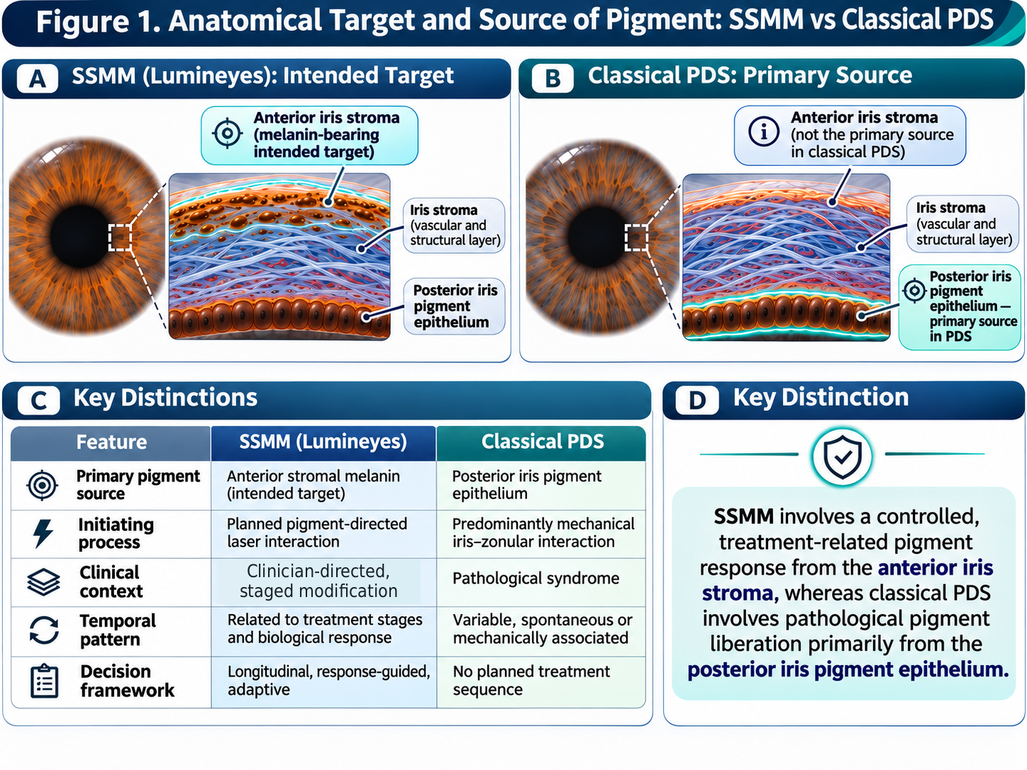
Figure 2
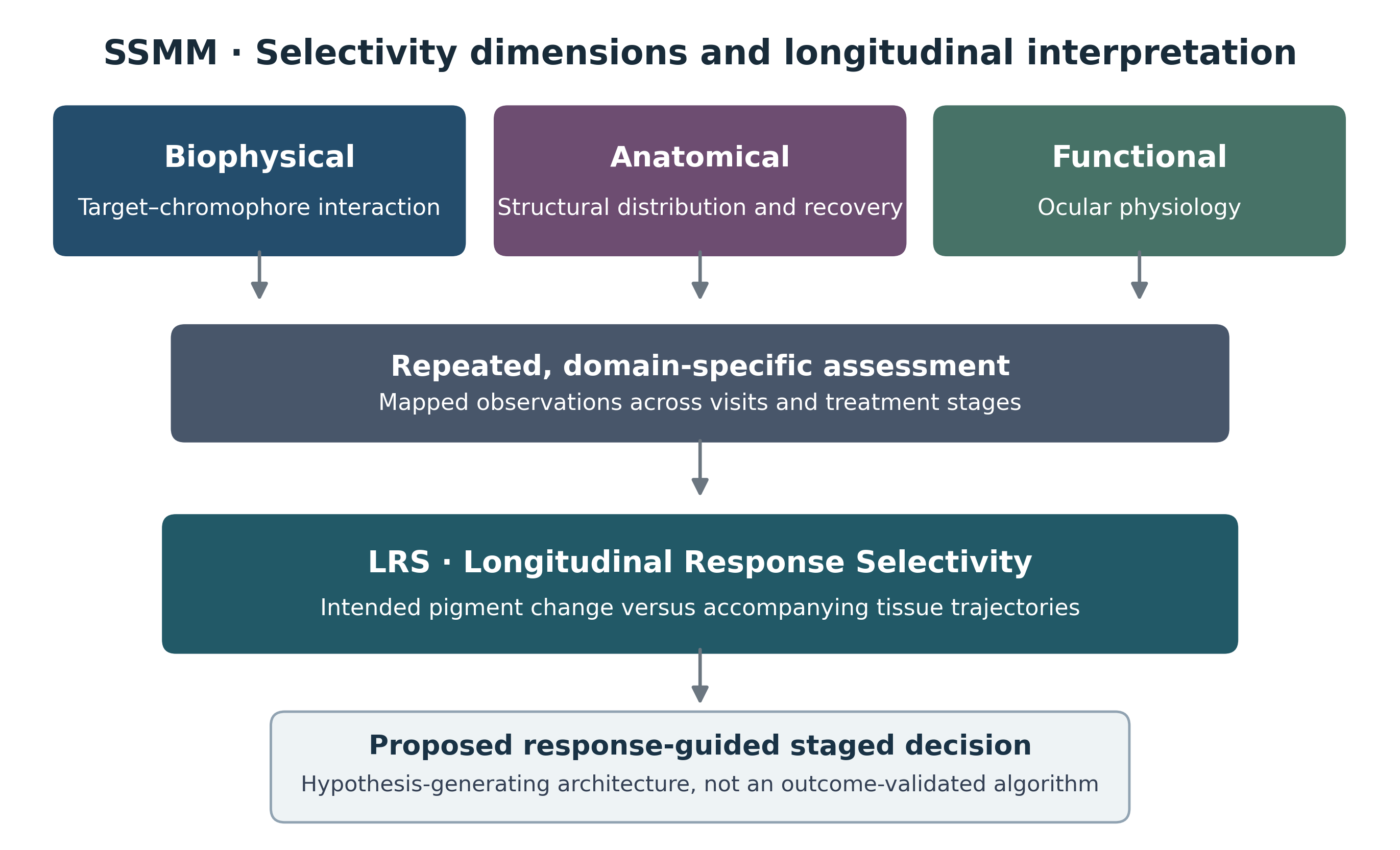
Figure 3