Full manuscript · Chapter 5 of 6
Temporal Response Architecture (TRA)
Associated research DOI: https://doi.org/10.5281/zenodo.23128049. Exact manuscript–record relationship awaits verification.
Temporal Response Architecture Following Laser-Based Iris Pigment Modification: An Integrative Mechanistic Review
Corresponding author: Mustafa Mete, MD
Abstract
Purpose: To organize biological responses after technically heterogeneous laser-based iris pigment modification into a coherent temporal framework and identify measurable variables for prospective investigation.
Methods: A focused mechanistic review integrated direct human procedural reports, preclinical iris studies, experimental trabecular and aqueous-outflow research, and studies of inflammatory, extracellular-matrix and cellular-clearance pathways. Evidence was classified by biological proximity to iris-directed laser exposure.
Results: Temporal Response Architecture (TRA) connects six overlapping domains: laser–tissue interaction and pigment modification; anterior-segment redistribution; inflammatory and cellular response; trabecular/outflow interaction; intraocular pressure (IOP) dynamics; and longitudinal resolution or progression. Clinical reports span protocol-specific stable IOP findings and marked pigment-associated ocular hypertension. Experimental evidence identifies candidate contributions from monocyte recruitment, MCP-1/CCR2 signaling, CD5L/AIM-associated phagocytosis, and extracellular-matrix regulation. Serial IOP, laser flare photometry, specular microscopy and anterior-segment optical coherence tomography provide complementary variables for prospective assessment.
Conclusion: TRA distinguishes elapsed time, observed ocular state and direction of change. Its integration of cellular mechanisms with multimodal longitudinal measurements supports testable research questions about recovery and persistent dysfunction within defined laser-exposure protocols.
Keywords: iris pigment modification; anterior segment; trabecular meshwork; aqueous outflow; intraocular pressure; phagocytosis
Introduction
Human iris appearance reflects pigmentation, stromal architecture, tissue thickness, light scattering, and other optical properties rather than pigment quantity alone. [1,2] Procedures intended to alter apparent eye color include corneal pigmentation, intraocular implants, and laser-directed alteration of iris pigmentation. [3] The laser literature remains procedurally heterogeneous, with reports using terms such as laser iris depigmentation, cosmetic iridoplasty, and iris color-change laser treatment despite substantial differences in wavelength, pulse characteristics, spot geometry, treatment density, treated area, number and spacing of exposures, postoperative medication, and surveillance. [4–11]
The post-laser response is often discussed as a single postoperative event. Biologically, however, laser–tissue interaction may initiate several related processes: pigment modification or mobilization, redistribution within the anterior segment, inflammatory activity, interaction with the conventional aqueous-outflow pathway, changes in IOP, cellular processing, and eventual resolution or persistence. These processes may overlap and interact rather than occur as discrete chronological stages, and the evidence informing them comes from different experimental settings.
This review integrates clinical and experimental evidence into a temporal model of anterior-segment response after iris-directed laser exposure. Temporal Response Architecture (TRA) connects the principal biological response domains and distinguishes elapsed time from observed ocular state and longitudinal trajectory. This structure provides a testable basis for serial clinical and mechanistic investigation.
Terminology and Treatment Architecture
Laser-based iris pigment modification is used here as an umbrella term for the diverse laser procedures reported in the literature. The abbreviation LID is retained when discussing publications whose authors specifically describe laser iris depigmentation. Exposure characteristics differ according to wavelength, pulse duration, spot geometry, fluence, treatment density, cumulative treated area, spacing of exposures, tissue characteristics, medication, and monitoring.
Within this review, Lumineyes™ denotes the selective, staged, response-guided framework for stromal melanin modulation. It is distinguished from other techniques within the broader laser iris depigmentation literature by its treatment architecture and clinical approach. Interpretations of clinical outcomes therefore remain specific to the protocol and exposure characteristics being studied.
Methods
A focused mechanistic review was conducted using PubMed records from database inception through 30 September 2026. Search domains covered iris depigmentation and pigment modification, pigment dispersion, trabecular meshwork and aqueous outflow, IOP, anterior-chamber inflammation, phagocytosis, and monocyte/macrophage recruitment. Targeted source verification and reference-list screening extended the synthesis to extracellular-matrix regulation, MCP-1/CCL2 signaling, CD5L/AIM-mediated phagocytosis, laser flare photometry, corneal endothelial imaging, and anterior-segment OCT. Reviews informed background, terminology, and citation chaining. [3,21,27,28,31–33] The review integrates findings narratively across biological domains rather than pooling outcomes from differing techniques.
Full search strategies, the study-level evidence map and contextual source classifications are provided in Online Resource 1.
Human studies and case reports were classified as direct procedural evidence when they described intentional laser interaction with iris pigmentation and reported findings relevant to predefined response domains. Experimental studies were classified as direct preclinical iris evidence when laser exposure was applied specifically to pigmented iris tissue. Non-iris studies were incorporated when they addressed mechanistically relevant components such as pigment–trabecular interaction, outflow regulation, laser-associated inflammatory signaling, cellular recruitment, phagocytosis, or particulate clearance. Discordant findings were retained when relevant, and the evidence was synthesized narratively across the predefined domains.
Evidence categories indicate biological proximity, not methodological quality: Category A, direct human procedural evidence; Category B, direct preclinical iris evidence; Category C, mechanistically analogous evidence; and Category D, integrative inference generated by the present synthesis.
Direct Evidence From Iris-Directed Laser Exposure
Direct evidence is limited and procedurally heterogeneous. In rabbit irides, Yildirim et al. reported histologic pigment reduction after frequency-doubled Nd:YAG exposure, with mild anterior-chamber cells/flare one day after treatment that resolved by the end of the first week and no significant IOP change within that protocol. [4] This demonstrates that iris-directed laser exposure can modify pigmentation and generate a transient anterior-segment response, but does not establish human kinetics.
Basoglu and Celik described 532-nm SLT-platform treatment for sectoral heterochromia using a gradual zonal approach. [5] Grimaldos Ruiz later reported a prospective series of 1176 eyes treated with a specific 532-nm Q-switched Nd:YAG protocol, with progressive color change, no statistically significant pre- versus post-treatment IOP difference in longitudinal analyses, and inflammatory findings in a subset. [6] These observations remain specific to the reported treatment architecture.
Conversely, case reports describe pigment dispersion, ocular hypertension, and secondary pigmentary glaucoma after other cosmetic iris-laser exposures. [7–11] Liu et al. described severe bilateral pigmentary glaucoma accompanied by paracentral acute middle maculopathy (PAMM), introducing a clinically relevant posterior-segment endpoint within the reported adverse-response phenotype. [10] These observations establish possible clinical response patterns and motivate mechanistic investigation of both anterior-segment dysfunction and its downstream consequences. Differences in exposure architecture, patient susceptibility, and surveillance remain essential to interpreting these reports.
Direct human reports are particularly informative for defining the range of possible response states, but their evidentiary roles differ. Case reports are useful for identifying adverse-response phenotypes and generating mechanistic questions; they cannot determine frequency. Larger protocol-specific series can characterize outcomes within the reported architecture, but do not establish equivalence with technically different approaches. TRA therefore uses both forms of evidence without treating either as a universal estimate of risk.
Mechanistic Response Domains
Target interaction is the first TRA domain. Because published protocols differ substantially, no single photodisruptive, photothermal, or photoablative mechanism should be assumed across the literature. TRA therefore uses the neutral sequence target interaction → pigment modification/mobilization. Visible iris lightening is not a quantitative surrogate for the amount of material entering the anterior segment: appearance also depends on stromal architecture and optical scattering, [1,2] and direct human evidence linking visible change to particulate burden is lacking.
Pigment-containing material may redistribute into the anterior chamber and angle after some procedures. [7–11] Clinical anterior-chamber cell grading cannot by itself determine whether observed particles represent inflammatory cells, pigment-containing material, or both. Preclinical iris evidence supports transient inflammation after laser exposure, [4] while SLT and laser-trabeculoplasty studies demonstrate early anterior-chamber activity, occasional transient pressure change, inflammatory cytokine signaling, and matrix-remodeling responses. [16,17] These models support plausibility but do not establish identical magnitude or timing after iris pigment modification.
The trabecular meshwork is biologically active rather than a passive filter, with phagocytic, contractile, and extracellular-matrix regulatory functions. [12,27,28] Experimental pigment exposure can reduce outflow facility and alter trabecular-cell behavior. Epstein et al. demonstrated acute outflow-facility reduction after pigment infusion in living monkeys followed by recovery and particulate uptake by trabecular cells and macrophages. [13] In experimental pigmentary-glaucoma systems, IOP elevation preceded reduced trabecular phagocytic activity, [14] while pigment exposure influenced migration, phagocytosis, cytoskeletal organization, and contractility. [15] These findings suggest a coupled mechanical–cellular response rather than passive particulate obstruction alone.
Extracellular-matrix (ECM) turnover provides a complementary regulatory axis. Laser-trabeculoplasty experiments link IL-1β and TNF-α signaling to matrix-metalloproteinase expression, [17] while studies of conventional outflow identify MMP-mediated ECM remodeling as part of pressure homeostasis. [27,28] In human trabecular-cell models, TGF-β2 and matrix stiffness can influence YAP/TAZ signaling, cytoskeletal organization, and contractile behavior. [29] These pathways provide candidate mechanisms through which inflammatory signaling and tissue biomechanics could influence recovery or persistent outflow resistance. Their relative contribution after specific iris-directed laser architectures remains an empirical question.
Experimental pigment may differ from laser-generated iris material in particle size, cellular association, tissue debris, melanin packaging, and inflammatory context. Post-laser pressure responses may therefore reflect interacting particulate, inflammatory, ECM-remodeling, pharmacological, tissue-injury, and host-susceptibility factors. A conceptual representation is R(t) = f(P, T, I, E, C, H; t), where P denotes particulate burden, T trabecular-cell function, I inflammatory signaling, E extracellular-matrix dynamics, C processing/clearance capacity, H host susceptibility, and t elapsed time. This notation organizes candidate variables rather than assigning numerical predictive weights.
Recovery can involve active cellular processing. Pigment models demonstrate particulate uptake during recovery of outflow function, [13] and SLT-related experiments document monocyte/macrophage recruitment to the trabecular meshwork and effects on conventional outflow. [18] MCP-1/CCL2 acts through the CCR2 pathway in experimental outflow systems; in perfused porcine eyes and Schlemm-canal endothelial-cell models, MCP-1 altered outflow facility and barrier-related measurements. [26] These observations establish a plausible chemokine-associated component of trabecular response, without equating SLT or experimental perfusion with iris pigment-modification procedures.
The CD5L/AIM–CD36 pathway adds a specific cellular-clearance mechanism. Nemoto et al. demonstrated AIM-enhanced phagocytosis in human trabecular-meshwork cells; in a mouse laser-iridotomy model, AIM deficiency delayed angle-debris clearance and recovery from transient ocular hypertension, whereas AIM treatment improved the experimental response. [22] TRA incorporates this evidence as a testable processing-capacity mechanism: a comparable post-iris-laser effect has not been established, but it provides a molecular candidate for investigating interindividual differences in clearance trajectories.
IOP is consequently treated in TRA as both a safety endpoint and a longitudinal response variable. A single measurement captures one state, whereas serial measurements can show stability, transient deviation, recovery, or persistent abnormality. Direct clinical reports illustrate this spectrum: some protocol-specific observations describe comparatively stable pressure behavior, [6] while case reports document substantial ocular hypertension and secondary glaucoma after other exposures. [7–11] The available literature cannot determine how much of this variability reflects treatment architecture, cumulative particulate burden, inflammation, baseline outflow susceptibility, medication, or procedural injury.
Temporal Organization and Proposed TRA
Available evidence does not support a universal post-exposure timetable. Relevant experimental systems show that inflammatory, trabecular, matrix-remodeling, and phagocytic responses evolve over time, [14–18,22,26–29] but their timing must be evaluated within each iris-directed laser protocol. TRA distinguishes elapsed time, ocular state, and trajectory: time describes chronology, state records the condition at an examination, and trajectory describes the direction and persistence of change across observations.
TRA organizes six partially overlapping domains: (1) laser–tissue interaction and pigment modification; (2) anterior-segment redistribution; (3) inflammatory and cellular response; (4) trabecular and aqueous-outflow interaction; (5) IOP and aqueous-dynamic response; and (6) longitudinal resolution or progression. ECM regulation, chemokine activity, and phagocytic clearance connect several domains. IOP may remain stable, deviate transiently, or show sustained alteration. Figure 1 illustrates the core architecture; candidate molecular and imaging measurements are specified in Table 1 and the prospective assessment strategy.
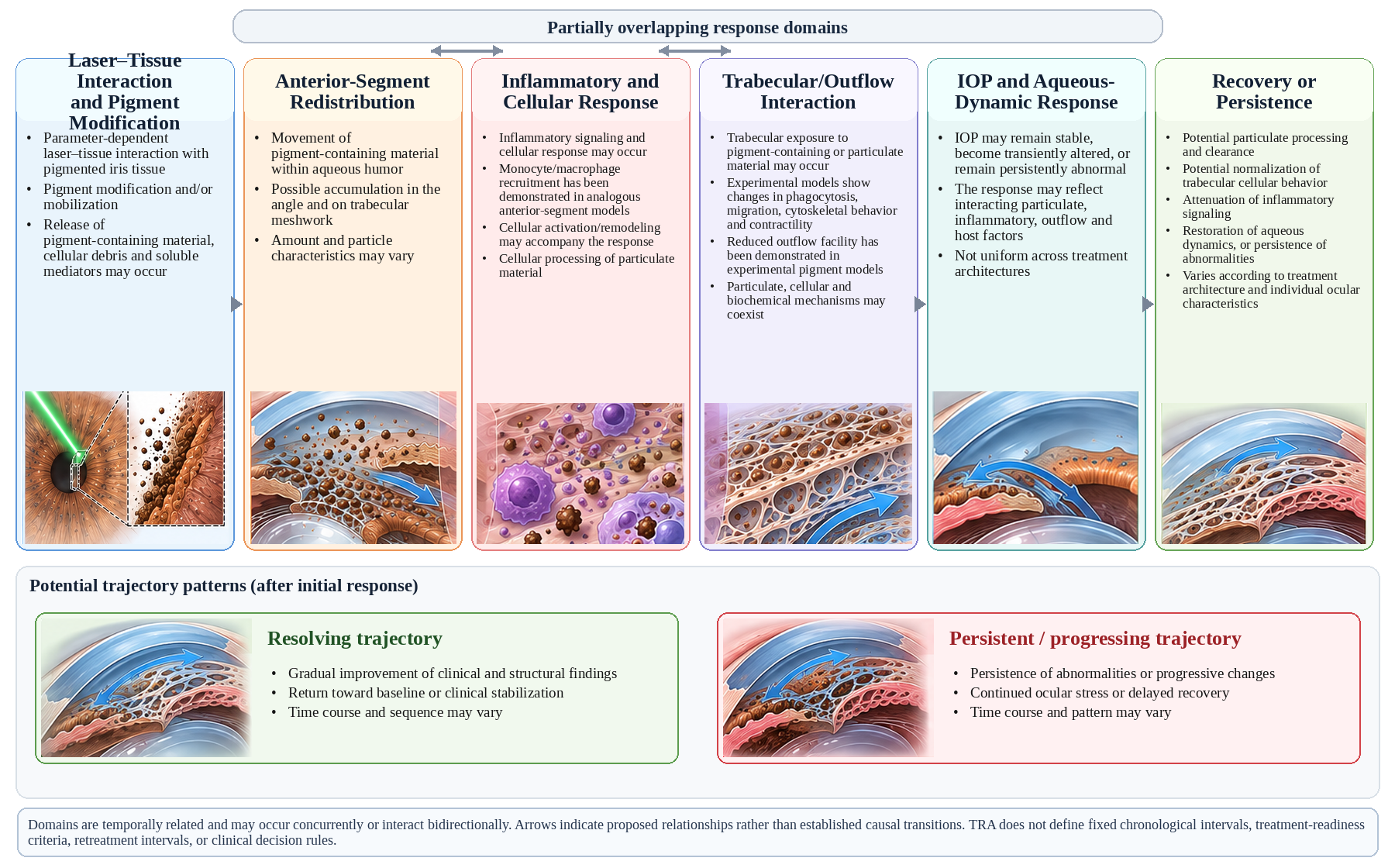
Fig. 1 Temporal Response Architecture following laser-based iris pigment modification. Initial laser–tissue interaction and pigment modification are linked with anterior-segment redistribution, inflammatory/cellular response, trabecular/outflow interactions, IOP/aqueous-dynamic response, and longitudinal resolving or persistent/progressing trajectories. Arrows express the proposed relationships among these interacting domains. ECM regulation and immune-cell recruitment connect domains; objective serial measurements described in the text provide a way to investigate their time-dependent behavior
Table 1 Evidence domains underlying Temporal Response Architecture
| TRA response domain | Representative evidence (category) | Mechanistic or measurement implication |
| Iris target interaction / pigment modification | Yildirim et al. [4]; Basoglu & Celik [5]; Grimaldos Ruiz [6] (A–B) | Laser–tissue interaction modifies iris pigmentation; exposure architecture governs the response. |
| Pigment redistribution / inflammatory response | Ning et al. [7]; Liu et al. [10]; Yildirim et al. [4]; De Keyser et al. [16] (A–C) | Pigment and cellular activity may enter the anterior chamber; laser flare photometry can quantify accompanying barrier response. [30,31] |
| Trabecular cellular interaction | Matsumoto & Johnson [12]; Dang et al. [14]; Wang et al. [15] (C) | Particle uptake, migration, phagocytosis, contractility, and cytoskeletal state influence the outflow response. |
| ECM remodeling and mechanotransduction | Bradley et al. [17]; De Groef et al. [27]; Keller & Peters [28]; Li et al. [29] (C) | MMP-associated turnover, TGF-β2 signaling, and matrix mechanics offer candidate links to outflow resistance. |
| Immune recruitment and cellular clearance | Alvarado et al. [18]; Tsuboi et al. [26]; Nemoto et al. [22] (C) | Monocyte recruitment, MCP-1/CCR2 activity and CD5L/AIM-mediated phagocytosis support active processing pathways. |
| IOP / aqueous dynamics | Grimaldos Ruiz [6]; Ong et al. [8]; Swampillai et al. [9]; Liu et al. [10] (A) | Clinical observations span stable pressure, transient changes and marked ocular hypertension in differing protocols. |
| Corneal and angle structural observations | Chaurasia & Vanathi [32]; Triolo et al. [33] (C) | Endothelial cell morphometry and AS-OCT angle/iris measures support standardized longitudinal characterization. |
| Posterior-segment outcome where indicated | Liu et al. [10]; Flores-Márquez et al. [24]; Wisdorf et al. [25] (A) | PAMM reported with severe IOP elevation and other retinal lesions motivate pathway-specific outcome assessment. |
| Longitudinal trajectory | Integrative TRA synthesis (D) | Serial direction and concordance across biological and clinical measures organize resolving versus persistent patterns. |
Table note: Evidence categories reflect biological proximity to the iris-laser question: A, direct human procedural evidence; B, direct preclinical iris evidence; C, mechanistically related biological or measurement evidence; D, integration by the present synthesis. Molecular and imaging methods are proposed for defined prospective designs; their use does not imply protocol-specific biological equivalence.
Discussion
TRA shifts interpretation from procedural naming toward biological exposure. Reports labeled as LID or cosmetic iris laser treatment may describe materially different exposures because pulse characteristics, beam geometry, density, cumulative treated area, staging, medication, and monitoring vary. Protocol-specific exposure characterization is consequently essential for interpreting the range of clinical findings, from stable pressure behavior to severe postoperative abnormalities.
TRA positions pigment burden within a wider system of outflow regulation. Experimental evidence demonstrates interactions among phagocytosis, monocyte recruitment, MCP-1/CCR2 signaling, CD5L/AIM-related clearance, cytoskeletal contractility, and ECM turnover. [13–18,22,26–29] Apparently similar pigment responses may produce different trajectories according to host outflow reserve, inflammatory context, matrix state, and processing capacity. This framework generates measurable hypotheses without treating any one pathway as the universal explanation for pressure change.
Longitudinal direction may add information beyond an isolated examination. IOP, anterior-chamber reaction, pigment, corneal status, and visible iris appearance remain essential observations, yet two eyes with the same finding at one visit may represent different biological states when one is resolving and the other is persistent or worsening. Prospective serial analysis can establish the incremental value of trajectory for characterizing recovery and adverse outcomes.
Posterior-segment observations merit a mechanistically differentiated extension of longitudinal surveillance. The PAMM finding reported alongside profound pigment-associated IOP elevation raises the possibility of a pressure/perfusion-related downstream pathway in certain adverse cases. [10] This route must be distinguished from direct or differently mediated laser-associated retinal injury described in other reports. [24,25] When clinically indicated, macular OCT and evaluation of visual function can document the relevant endpoint without expanding the central TRA model into a universal retinal injury mechanism.
This distinction is particularly relevant to staged treatment. A response-guided architecture uses the evolving ocular state alongside elapsed time. The selective, staged Lumineyes™ framework provides the clinical context for this approach. L-SAFE addresses observable anterior-segment safety state and its direction of change, [19] while TRA maps underlying biological response domains. L-OAT separates visible phenotype, iris architecture, and OCT-derived signal for quantitative phenotyping. [20] These complementary perspectives distinguish treatment exposure, biological response, clinical state, and structural phenotype.
TRA brings established biological processes into a single evidence-organized anterior-segment architecture. It links direct human findings, preclinical iris evidence, mechanistic analogues, and integrative inference without conflating their respective evidentiary roles. Its contribution lies in connecting laser–tissue interaction, subsequent pigment and outflow biology, and longitudinal clinical trajectory within a structure that can be investigated prospectively.
Research Implications and Limitations
Prospective evaluation should combine serial clinical, functional, and structural measurements in the same eye. The core observations include IOP, slit-lamp grading of anterior-chamber cells and pigment, gonioscopy, standardized iris photography, and corneal examination. Complementary noninvasive tools can quantify distinct domains: laser flare photometry for blood–aqueous barrier activity, specular microscopy for endothelial structure, and anterior-segment OCT for iris and angle morphology. [30–33] The observation schedule should be predefined by study design and aligned with the exposure protocol, with the same instruments and acquisition methods used for longitudinal comparisons.
Laser flare photometry quantifies aqueous light scatter associated with blood–aqueous barrier disturbance in photon counts per millisecond and can detect differences not fully represented by slit-lamp flare grades. [30,31] It offers an objective inflammatory-response variable for intra-eye trend analysis; clinical interpretation also depends on baseline values, optical clarity, device characteristics, and repeated-measure precision. Universal flare thresholds for retreatment readiness or for assigning TRA trajectories have not been established.
Specular microscopy provides endothelial cell density, mean cell area, coefficient of variation in cell size, and hexagonality, while pachymetry and slit-lamp findings provide complementary corneal context. [32] Serial analysis should document acquisition quality, the cells sampled, central or peripheral location where available, and device/method consistency. Anterior-segment OCT can quantify iris thickness or curvature and angle parameters such as angle-opening distance and trabecular–iris space area; these variables characterize anatomy but do not directly measure pigment clearance or outflow function. [33]
A proposed prospective dataset can therefore align: exposure parameters and staging; baseline phenotype and angle anatomy; repeated IOP and inflammatory measurements; iris and angle imaging; endothelial morphometry; and clinically indicated macular or visual-function observations. Resolving trajectories would be investigated through reproducible stabilization or return toward baseline across relevant variables; persistent/progressing trajectories through sustained or worsening abnormalities. Biomarker combinations and their predictive utility should be derived from serial data rather than assigned fixed numerical cutoffs.
Priority research questions include the temporal relation among particulate burden, barrier response, ECM and immune signaling, corneal or angle structural change, and IOP; the reproducibility of multivariable patterns within well-defined treatment architectures; and whether direction of change adds information beyond isolated measurements. Laser-generated iris material requires improved characterization, and no single imaging or molecular measure currently defines completion of biological recovery. These gaps directly motivate prospective repeated-measures designs.
The principal interpretive constraint is heterogeneity of direct human evidence and the need to distinguish iris-directed exposures from pigment infusion, SLT, laser-iridotomy, and glaucoma-derived cellular models. Individual case reports characterize possible clinical phenotypes but not incidence. Laser-associated maculopathy, [24] traumatic lamellar injury, [25] and iris perforation [23] illustrate additional potential endpoints whose pathways may differ from, or in selected cases be linked downstream to, an anterior-segment pressure response. TRA keeps these routes explicit while centering its mechanistic analysis on pigment, inflammation, trabecular function, and longitudinal ocular state.
Conclusion
Temporal Response Architecture integrates laser–tissue interaction, pigment redistribution, inflammatory signaling, trabecular and extracellular-matrix regulation, cellular clearance, aqueous-outflow behavior, and longitudinal pressure response into a connected mechanistic model. Molecular evidence for MCP-1/CCR2 signaling, MMP-associated remodeling, and CD5L/AIM-mediated phagocytic processing identifies new candidate pathways for mechanistic study. A coordinated prospective approach using serial IOP, inflammatory measurements, specular microscopy, and anterior-segment imaging can test how these processes relate to resolving or persistent response trajectories within defined laser exposure protocols.
References
1. Imesch PD, Wallow IHL, Albert DM. The color of the human eye: a review of morphologic correlates and of some conditions that affect iridial pigmentation. Surv Ophthalmol. 1997;41(Suppl 2):S117–S123. https://doi.org/10.1016/S0039-6257(97)80018-5
2. Sturm RA, Larsson M. Genetics of human iris colour and patterns. Pigment Cell Melanoma Res. 2009;22(5):544–562. https://doi.org/10.1111/j.1755-148X.2009.00606.x
3. D’Oria F, Abu-Mustafa SK, Alio JL. Cosmetic change of the apparent color of the eye: a review on surgical alternatives, outcomes and complications. Ophthalmol Ther. 2022;11(2):465–477. https://doi.org/10.1007/s40123-022-00458-2
4. Yildirim Y, Duzgun E, Kar T, et al. Evaluation of color-changing effect and complications after Nd:YAG laser application on iris surface. Med Sci Monit. 2016;22:107–114. https://doi.org/10.12659/MSM.895086
5. Basoglu A, Celik U. The effect of SLT laser application on iris to treat sectorial heterochromia: a promising technique. Eye Contact Lens. 2018;44(Suppl 1):S352–S354. https://doi.org/10.1097/ICL.0000000000000374
6. Grimaldos Ruiz P. Photoablative cosmetic iridoplasty: effective, safe, and predictable—eye color change in 1176 eyes. Int Ophthalmol. 2021;41(4):1381–1393. https://doi.org/10.1007/s10792-021-01693-5
7. Ning B, Baboolal S, Gizzi C, Nolan W. A case of secondary pigment dispersion following laser to cosmetically lighten the irises. J Glaucoma. 2022;31(2):133–135. https://doi.org/10.1097/IJG.0000000000001790
8. Ong AY, Ching-A-Sue G, Krilis M, Guirao-Navarro MC, Hornby S, Jutley G. Refractory iatrogenic pigmentary glaucoma secondary to cosmetic laser treatment: a case report. Eur J Ophthalmol. 2023;33(2):NP1–NP4. https://doi.org/10.1177/11206721211050338
9. Swampillai AJ, Sherman T, Garg A, Tan IJ, Lim KS. Secondary pigmentary glaucoma following cosmetic laser treatment to alter iris colour. Cont Lens Anterior Eye. 2023;46(2):101754. https://doi.org/10.1016/j.clae.2022.101754
10. Liu J, Korban S, Moster MR, Rhéaume MA, Wang Q. Bilateral severe iatrogenic pigmentary glaucoma following laser treatment for cosmetic iris color change. Am J Ophthalmol Case Rep. 2023;32:101927. https://doi.org/10.1016/j.ajoc.2023.101927
11. Jaber W, Nemet M, Waisbourd M. Laser iris depigmentation resulting in markedly elevated intraocular pressure and herpes simplex keratitis with corneal scarring: case report. Case Rep Ophthalmol. 2026;17(1):312–317. https://doi.org/10.1159/000551226
12. Matsumoto Y, Johnson DH. Trabecular meshwork phagocytosis in glaucomatous eyes. Ophthalmologica. 1997;211(3):147–152. https://doi.org/10.1159/000310782
13. Epstein DL, Freddo TF, Anderson PJ, Patterson MM, Bassett-Chu S. Experimental obstruction to aqueous outflow by pigment particles in living monkeys. Invest Ophthalmol Vis Sci. 1986;27(3):387–395.
14. Dang Y, Waxman S, Wang C, et al. Intraocular pressure elevation precedes a phagocytosis decline in a model of pigmentary glaucoma. F1000Res. 2018;7:174. https://doi.org/10.12688/f1000research.13797.2
15. Wang C, Dang Y, Loewen RT, et al. Impact of pigment dispersion on trabecular meshwork cells. Graefes Arch Clin Exp Ophthalmol. 2019;257(6):1217–1230. https://doi.org/10.1007/s00417-019-04300-7
16. De Keyser M, De Belder M, De Groot V. Randomized prospective study of the use of anti-inflammatory drops after selective laser trabeculoplasty. J Glaucoma. 2017;26(2):e22–e29. https://doi.org/10.1097/IJG.0000000000000522
17. Bradley JM, Anderssohn AM, Colvis CM, et al. Mediation of laser trabeculoplasty-induced matrix metalloproteinase expression by IL-1β and TNFα. Invest Ophthalmol Vis Sci. 2000;41(2):422–430.
18. Alvarado JA, Katz LJ, Trivedi S, Shifera AS. Monocyte modulation of aqueous outflow and recruitment to the trabecular meshwork following selective laser trabeculoplasty. Arch Ophthalmol. 2010;128(6):731–737. https://doi.org/10.1001/archophthalmol.2010.85
19. Mete M. Lumineyes Anterior-Segment Safety Staging (L-SAFE): A Proposed Framework for Pre-Exposure Assessment in Laser Iris Pigment Reduction. Zenodo. 2026. https://doi.org/10.5281/zenodo.23046455
20. Mete M. Lumineyes Optical Architectural Typology (L-OAT): a multimodal hypothesis for quantitative iris phenotyping. Zenodo. 2026. https://doi.org/10.5281/zenodo.23066772
21. D’Oria F, Alio JL. Surgical techniques for cosmetic eye color change: a narrative review. Ophthalmol Ther. 2025;14(8):1685–1694. https://doi.org/10.1007/s40123-025-01177-0
22. Nemoto H, Honjo M, Arai S, Miyazaki T, Aihara M. Apoptosis inhibitor of macrophages/CD5L enhances phagocytosis in the trabecular meshwork cells and regulates ocular hypertension. J Cell Physiol. 2023;238(10):2451–2467. https://doi.org/10.1002/jcp.31097
23. Ferrari F. Case report of an iris perforation after YAG laser iris depigmentation for aesthetic reasons. J Fr Ophtalmol. 2021;44(5):e283–e285. https://doi.org/10.1016/j.jfo.2020.07.016
24. Flores-Márquez A, Moreno-Gutiérrez JA, Chinchurreta-Capote A, et al. Laser-induced maculopathy after iris depigmentation cosmetic treatment. Can J Ophthalmol. 2023;58(1):e29–e31. https://doi.org/10.1016/j.jcjo.2022.05.012
25. Wisdorf K, Hami H, Widder RA, Rössler GF, Kiessling D. Traumatic lamellar macular hole and epiretinal gliosis after cosmetic laser iridoplasty. Ophthalmologie. 2025;122(4):308–311. https://doi.org/10.1007/s00347-025-02184-z
26. Tsuboi N, Inoue T, Kawai M, et al. The effect of monocyte chemoattractant protein-1/CC chemokine ligand 2 on aqueous humor outflow facility. Invest Ophthalmol Vis Sci. 2012;53(10):6702–6707. https://doi.org/10.1167/iovs.12-10376
27. De Groef L, Van Hove I, Dekeyster E, Stalmans I, Moons L. MMPs in the trabecular meshwork: promising targets for future glaucoma therapies? Invest Ophthalmol Vis Sci. 2013;54(12):7756–7763. https://doi.org/10.1167/iovs.13-13088
28. Keller KE, Peters DM. Pathogenesis of glaucoma: extracellular matrix dysfunction in the trabecular meshwork—a review. Clin Exp Ophthalmol. 2022;50(2):163–182. https://doi.org/10.1111/ceo.14027
29. Li H, Raghunathan VK, Stamer WD, Ganapathy PS, Herberg S. Extracellular matrix stiffness and TGFβ2 regulate YAP/TAZ activity in human trabecular meshwork cells. Front Cell Dev Biol. 2022;10:844342. https://doi.org/10.3389/fcell.2022.844342
30. Tugal-Tutkun I, Herbort CP. Laser flare photometry: a noninvasive, objective, and quantitative method to measure intraocular inflammation. Int Ophthalmol. 2010;30(5):453–464. https://doi.org/10.1007/s10792-009-9310-2
31. Swampillai AJ, Nigam C, Dowse E, Lim KS. Quantification of postoperative inflammation in laser and surgical procedures for glaucoma with laser flare photometry: a review of the literature. Curr Eye Res. 2024;49(3):225–234. https://doi.org/10.1080/02713683.2023.2282940
32. Chaurasia S, Vanathi M. Specular microscopy in clinical practice. Indian J Ophthalmol. 2021;69(3):517–524. https://doi.org/10.4103/ijo.IJO_574_20
33. Triolo G, Barboni P, Savini G, et al. The use of anterior-segment optical-coherence tomography for the assessment of the iridocorneal angle and its alterations: update and current evidence. J Clin Med. 2021;10(2):231. https://doi.org/10.3390/jcm10020231
Statements and Declarations
Funding: No external funding was received for this work.
Competing interests: Mustafa Mete is the developer of the Lumineyes™ framework discussed in this article.
Author contributions: Mustafa Mete: conceptualization, methodology, investigation, literature synthesis, visualization, writing – original draft, writing – review and editing, and final manuscript approval.
Ethics approval: Not applicable; this article is a literature-based review and does not report new research involving human participants or animals.
Consent to participate and publication: Not applicable.
Data availability: No new datasets were generated or analyzed. The sources used in this review are identified in the reference list and Online Resource 1.
Supplementary Information
Online Resource 1: PubMed search strategies, study-level evidence mapping and contextual source classifications (ESM_1.pdf).