Laser Iris Depigmentation for Eye-Color Change: Stromal Pigment Biology, Anterior-Segment Response, and Longitudinal Clinical Assessment
Mustafa Mete · Preprints.org · Posted 11 November 2026 · CC BY 4.0 · Not peer-reviewed.
Official source DOI: 10.20944/preprints202611.1201.v1 · Download the source PDF · Compendium contents
Source page 1
Review Laser Iris Depigmentation for Eye-Color Change: Stromal Pigment Biology, Anterior-Segment Response, and Longitudinal Clinical Assessment
Mustafa Mete
MyLumineyes, Tunek Sokak No: 5/13, Göztepe, Istanbul 34730, Türkiye; info@mylumineyes.com
Abstract
Laser iris depigmentation (LID) modifies visible iris color through interaction with pre-existing anterior stromal pigmentation. Its biological effects extend beyond the immediate optical result to pigment-bearing cells, extracellular pigment material, and potentially the aqueous outflow pathway. Selective photothermolysis provides a physical basis for pigment-targeted pulsed irradiation. Histological studies in pigmented rabbit eyes document reduced anterior stromal pigmentation and an increased melanophage response after laser exposure. Pigment-perfusion experiments and trabecular-cell studies identify mechanisms through which pigment burden and cellular processing can influence outflow physiology. Human literature includes a 1,176-eye single-program clinical series and independent reports of secondary pigment dispersion and pigmentary glaucoma. This narrative review integrates iris morphology, laser–tissue interaction, treatment-related pigment dispersion, anterior-segment physiology, sequential clinical assessment, and long-term optical outcomes. It proposes controlled pigment dispersion as an observational construct: pigment liberation is assessed by its distribution, persistence, associated tissue response, and clinical consequences rather than treated as a binary event. A longitudinal response-guided framework connects these biological domains to follow-up, patient selection, and decisions about further exposure. Clinical evaluation rests on the combined assessment of iris appearance, anterior-segment status, pressure, and visual function.
Keywords: laser iris depigmentation; iris stroma; eye color change; melanin; pigment dispersion; trabecular meshwork; intraocular pressure; longitudinal monitoring
1. Introduction
Cosmetic alteration of eye color can be achieved by changing the pigment of the native iris, placing pigment within the cornea, or implanting a colored device. These interventions share a visible objective but operate in different anatomical compartments. Laser iris depigmentation (LID) acts on pre-existing iris pigmentation and therefore requires interpretation of the response of living uveal tissue, not simply assessment of an optical result. [1,2,3] The clinical relevance of LID follows from a linked biological sequence: optical absorption by pigment, laser-induced alteration of pigment-bearing stromal cells, movement or processing of pigment-derived material, and potential effects within the anterior chamber and aqueous outflow pathway. Selective photothermolysis establishes the fundamental relationship between chromophore absorption, pulse duration, and localized tissue response; nanosecond Q-switched experiments further demonstrate wavelength-dependent melanosome disruption. [4,5] Direct iris laser experiments connect these principles to stromal pigmentation change and cellular processing. [6] Eye color also depends on the remaining stromal optical architecture, so the visible result and anteriorsegment physiological response should be evaluated as distinct, linked outcomes. [1,2] This narrative review synthesizes four complementary evidence streams: iris pigmentation morphology, direct experimental iris laser exposure, pigment and trabecular outflow physiology, and
Source page 2
clinical outcomes after elective eye-color procedures. Relevant PubMed/MEDLINE literature and reference lists were screened through November 2026, emphasizing experimental studies, clinical series, complication reports, and relevant reviews. The central proposition is that visible pigmentation change and anterior-segment tissue response are connected but noninterchangeable endpoints. The resulting framework is organized around biological compartments, sequential observations, and procedure-specific evidence.
2. The Iris as a Pigmented Optical Tissue
2.1. Melanin Distribution and Iris Color
Normal variation in iris color is strongly associated with melanin granule content and its distribution within the anterior iris. Morphological studies indicate that differences between light and dark irides largely reflect the amount of melanin contained in stromal melanocytes, rather than a corresponding difference in their total number. The posterior iris pigment epithelium remains densely pigmented across ordinary iris colors. Visible appearance results from absorption by melanin combined with wavelength-dependent scattering and transmission in stromal tissue. [1,2] This anatomy has a direct consequence for depigmentation: reducing superficial melanin changes the optical contribution of deeper stromal components without creating a new iris pigment. Stromal fiber organization, residual pigment distribution, and local tissue geometry influence brightness and hue. Pigment reduction and optical appearance are linked outcomes, but they are not equivalent measurements. [1,2]
2.2. Tissue Architecture as a Determinant of Response
The iris stroma contains melanocytes, fibroblasts, collagen-rich extracellular matrix, vessels, nerves, immune-related cells, and smooth-muscle structures arranged in a functional anteriorsegment tissue. Pigment-bearing cells are therefore embedded within tissue that also contributes to pupillary and optical behavior. The spatial relationship between pigment, stromal matrix, and vascular elements is central to interpreting localized laser exposure. [1,6] Anatomical selectivity, pigment response, and preservation of tissue function are distinct questions. Color photography can document visible lightening but cannot, by itself, establish the condition of stromal cells, collagen architecture, angle pigmentation, or endothelial integrity. These endpoints require corresponding clinical or structural observations.
3. Experimental Basis of Laser-Induced Pigment Modification
3.1. Selective Photothermolysis and Melanin-Targeted Pulsed Irradiation
Selective photothermolysis explains how a pulsed laser can preferentially affect an absorbing chromophore through the combined influence of wavelength-dependent absorption, pulse duration, and the thermal properties and dimensions of the target. Anderson and Parrish established that selective injury can be localized to pigmented cellular structures and organelles when the optical exposure is matched to their physical properties. This principle supplies the biophysical starting point for pigment-directed laser procedures. [4] Subsequent Q-switched Nd:YAG experiments demonstrated disruption of melanosomes in pigmented skin with nanosecond-domain exposures at 355, 532, and 1064 nm, together with wavelength-dependent tissue responses. These observations connect selective light absorption to microscopic pigment alteration. In the iris, the same general laser–chromophore principles operate within a different tissue architecture: pigmented stromal cells are embedded among collagen, vascular elements, and functional iris structures. Direct iris experiments therefore provide the tissuespecific bridge between the established optical principle and observed pigment and cellular changes. [5,6]
Source page 3
3.2. Direct Iris Evidence
Yildirim and colleagues examined 532-nm frequency-doubled Nd:YAG exposure in 12 pigmented rabbit eyes over three sessions. Histological assessment at day 60 demonstrated reduction of anterior stromal pigment density and an increase in melanophages, with greater pigment reduction in the higher-exposure group. The investigators also described mild transient anteriorchamber inflammation; iris color change became more evident during follow-up. This experiment connects visible depigmentation with changes in pigment-bearing cells and local pigment-processing activity. [6] The histological findings are particularly informative because they distinguish immediate laser– pigment interaction from subsequent cellular behavior. Reduction in pigmented stromal cells, melanophage accumulation, and delayed evolution of visible appearance can occur within the same treatment sequence. The observations support a dynamic tissue-response model rather than an instantaneous optical endpoint. [6]
3.3. Pigment Liberation, Redistribution, and Cellular Processing
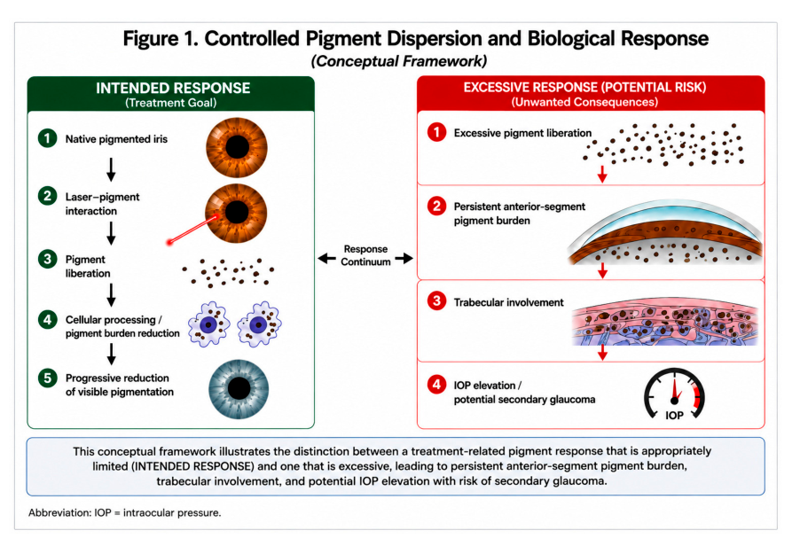
Laser-induced pigment change can produce at least two related processes: alteration of the anterior stromal pigment distribution and generation of pigment-derived material that may remain locally or enter the anterior chamber. The rabbit histology directly establishes stromal melanophage involvement after iris exposure; the extent of material transported into the human drainage pathway is a separate measurement question. The biological pathway is best examined as a sequence of compartments: treated iris stroma, aqueous environment, and outflow tissues. [6,7] Figure 1 integrates these compartments. It separates the optical result—appearance of the treated iris—from physiological responses such as pigment deposition, cellular handling, and changes in aqueous outflow. This distinction makes it possible to describe an effective cosmetic response and an unfavorable anterior-segment response using different outcome measures.
Figure 1. Controlled pigment dispersion and the biological response continuum. The left panel represents the
intended sequence of pigment alteration and processing; the right depicts excessive or persistent pigment
burden and potential outflow consequences. The image is a conceptual illustration, not a demonstration that all
treated irides evolve to blue or that a numerical safe pigment threshold has been established. [6,7,8].
Source page 4
3.4. Structural Response and Visible Outcome
The direct rabbit evidence includes a contrast between the time of laser exposure and the later appearance of the iris. Localized pigment accumulation and patchiness were described early in the observation period, with more evident lightening by the final day-60 assessment. Histology showed altered anterior stromal pigmentation and melanophages. This temporal sequence makes the postexposure state a moving biological target: an image recorded at one visit captures a particular stage of response rather than the complete treatment outcome. [6] Stromal integrity is consequently an independent endpoint. The presence of reduced surface pigment establishes an effect on pigmentation, whereas confirmation of intact pupillary function, normal ocular pressure, and absence of persistent inflammation requires separate observation. Experimental histology supplies one layer of anatomical information; longitudinal clinical assessment supplies functional information. A rigorous clinical account reports these domains separately rather than defining success solely by the amount of visible color change. [6,9]
3.5. Controlled Pigment Dispersion: A Treatment-Response Construct
Controlled pigment dispersion is used here to describe the intended, clinically observed sequence of pigment liberation, redistribution or cellular processing, and subsequent change in anterior-segment pigment burden during staged iris depigmentation. The term refers to how the response is evaluated over time. It is distinct from primary pigment dispersion syndrome, in which the initiating pathology is typically iris–zonular mechanical interaction, and from secondary pigmentary glaucoma, in which pigment-associated outflow dysfunction has become clinically consequential. [6,10] The distinction has four measurable dimensions: the amount and distribution of pigment released; its persistence in the aqueous and angle; the accompanying inflammatory or tissue response; and the trajectory of aqueous outflow function, clinically assessed principally through IOP and angle examination. Pigment liberation may accompany the desired iris color change, while an excessive or sustained burden may threaten trabecular function. These responses form a continuum rather than two biologically isolated categories. [7,8,10] Within this construct, the word “controlled” denotes exposure that is staged and monitored with decisions informed by observed physiology. It does not denote an experimentally established safe pigment concentration. A single normal pressure reading and a favorable photograph represent different observations, neither of which substitutes for a longitudinal assessment. Figure 1 summarizes the intended and excessive ends of the proposed response continuum. [7,10]
4. Pigment Burden and the Aqueous Outflow Pathway
4.1. Pigment Deposition Is a Physiological Challenge
Pigment dispersion syndrome and pigmentary glaucoma establish the clinical significance of pigment accumulation in the anterior-chamber angle. Their underlying anatomic mechanism—often involving iris–zonular contact—is different from treatment-related pigment release. Both settings nevertheless bring pigment into contact with the trabecular meshwork, where outflow resistance may change. [10] In living monkeys, Epstein and colleagues infused pigment particles into the anterior chamber and observed an acute decrease in outflow facility, followed by recovery toward baseline at the next weekly measurement. Ultrastructural examination demonstrated particle uptake by trabecular endothelial cells and macrophages. The experiment shows that an early pigment-associated reduction in outflow facility may coexist with subsequent cellular clearance and physiological recovery; cumulative pigment deposition alone does not fully explain persistent human pigmentary glaucoma. [7]
Source page 5
4.2. Trabecular Cells Respond to More Than Particle Quantity
Wang and colleagues studied porcine trabecular meshwork cells exposed to pigment. Pigment loading reduced phagocytosis and migration, increased stress-fiber formation and cell contraction, and involved Rho-associated signaling. The findings establish cellular routes through which exposure to pigment can alter outflow-tissue behavior. They also distinguish the physical presence of particles from the functional response of tissue handling them. [8] Additional mechanistic context comes from selective laser trabeculoplasty (SLT), where Alvarado and colleagues documented monocyte recruitment to the trabecular meshwork and demonstrated increased outflow facility or endothelial conductivity in experimental systems. These observations identify immune-cell participation in outflow regulation. SLT directly targets the drainage tissue, whereas LID targets the iris; the studies therefore illuminate common cellular capabilities without establishing an identical treatment mechanism. [11] Taken together, the primate perfusion, trabecular-cell, and SLT findings support a more informative physiological formulation: the clinical effect of liberated pigment depends on exposure burden, persistence, tissue handling, and the functional response of the outflow pathway. These dimensions are relevant to monitoring during staged iris treatment. [7,8,11]
4.3. Pressure Trajectory and Pigment-Processing Capacity
The distinction between transient and sustained pressure change matters. An acute rise may accompany a sudden pigment challenge, whereas persistent outflow impairment implies an ongoing disturbance in drainage physiology. The primate experiments show that outflow can recover as particles are handled by resident cells; pigment-treated trabecular cultures show that phagocytic capability and cytoskeletal state can themselves change under pigment exposure. Both findings make longitudinal pressure measurements and anterior-segment examinations more informative than a single value obtained at one postoperative visit. [7,8] In clinical follow-up, the pressure curve should be interpreted with anterior-chamber pigmentation and inflammation, angle findings, ocular symptoms, and structural glaucoma assessment when indicated. These are complementary observations: pressure reflects an integrated physiological state, whereas pigment and inflammatory findings describe aspects of the potential tissue challenge. The two need not change simultaneously. Reports of severe pigmentary glaucoma after cosmetic iris laser procedures illustrate the importance of this combined interpretation. [10,12,13]
4.4. From Particle Load to Time-Dependent Outflow Response
Aqueous outflow response can be understood as an interaction between the pigment challenge and the tissue’s evolving capacity to handle that challenge. In the monkey perfusion experiments, outflow facility initially decreased sharply after intracameral pigment administration and subsequently approached baseline during follow-up. In porcine trabecular cultures, pigment loading changed phagocytic activity, migration, and cytoskeletal behavior. These results are complementary: a physiological change can evolve over time while the cells responsible for processing particles are themselves affected by the exposure. [7,8] Temporal Response Architecture (TRA) extends this interpretation across successive observation points by distinguishing elapsed time after exposure, the ocular state at examination, and the direction of change between visits. Pigment mobilization, cellular activity, angle findings, inflammation, and IOP are thereby treated as related longitudinal response domains rather than as events assumed to follow a universal timetable. This structure makes it possible to compare early and delayed observations within the same eye and across treatment stages. [14] The monocyte recruitment reported after selective laser trabeculoplasty adds a distinct example of immune-cell participation in outflow regulation. Because SLT acts at the trabecular meshwork rather than at the iris, it is mechanistic context, not a demonstration of the same cascade following
Source page 6
cosmetic LID. Taken together, the studies define hypotheses for direct investigation of pigment particle characteristics, cellular clearance, angle findings, and serial IOP following iris treatment. [7,8,11]
5. Human Evidence: Effectiveness, Complications, and Interpretation
5.1. Prospective Procedural Outcomes
Grimaldos Ruiz published a prospective photoablative cosmetic iridoplasty series comprising 1,176 treated eyes of 588 patients from January 2017 to February 2020. Within that program, the mean recorded IOP was 10.6 mmHg before and 10.8 mmHg after treatment, without a statistically significant group difference (reported p = 0.63204); corrected visual acuity was also reported as stable overall. Delayed, brief iritis was described in approximately 25% of patients, and patient satisfaction at the end of treatment was reported as 95%. The publicationʹs nine-year period refers to the broader investigative program beginning in 2012; the 1,176-eye treatment cohort itself was enrolled during 2017–2020 and should not be presented as a cohort in which every eye completed nine years of observation. These data describe a specific procedure, patient selection process, and ascertainment strategy. [9]
5.2. Documented Adverse Clinical Phenotypes
Independent clinical reports identify a contrasting phenotype characterized by substantial pigment release, marked angle pigmentation, and pressure elevation. Ning and colleagues described bilateral secondary pigment dispersion after cosmetic iris laser exposure. Swampillai and colleagues reported secondary pigmentary glaucoma. Ong and colleagues described bilateral glaucoma with intense angle pigmentation and pressure elevation requiring trabeculectomy. These observations identify the anterior chamber and trabecular outflow pathway as clinically consequential sites of response. [13,15,16] Liu and colleagues reported a patient with intraocular pressures of 50 and 42 mmHg after four bilateral cosmetic laser sessions, accompanied by 4+ floating pigmented cells and severe visual consequences. In a 2026 two-patient case report, Jaber and colleagues documented marked pressure elevations (34 mmHg bilaterally in one patient; 60 mmHg in one eye of the other). One patient subsequently developed herpes simplex keratitis and corneal scarring; the report establishes the clinical sequence rather than a proven laser-specific cause of the infection. [12,17]
5.3. Reading the Evidence Without Collapsing Study Designs
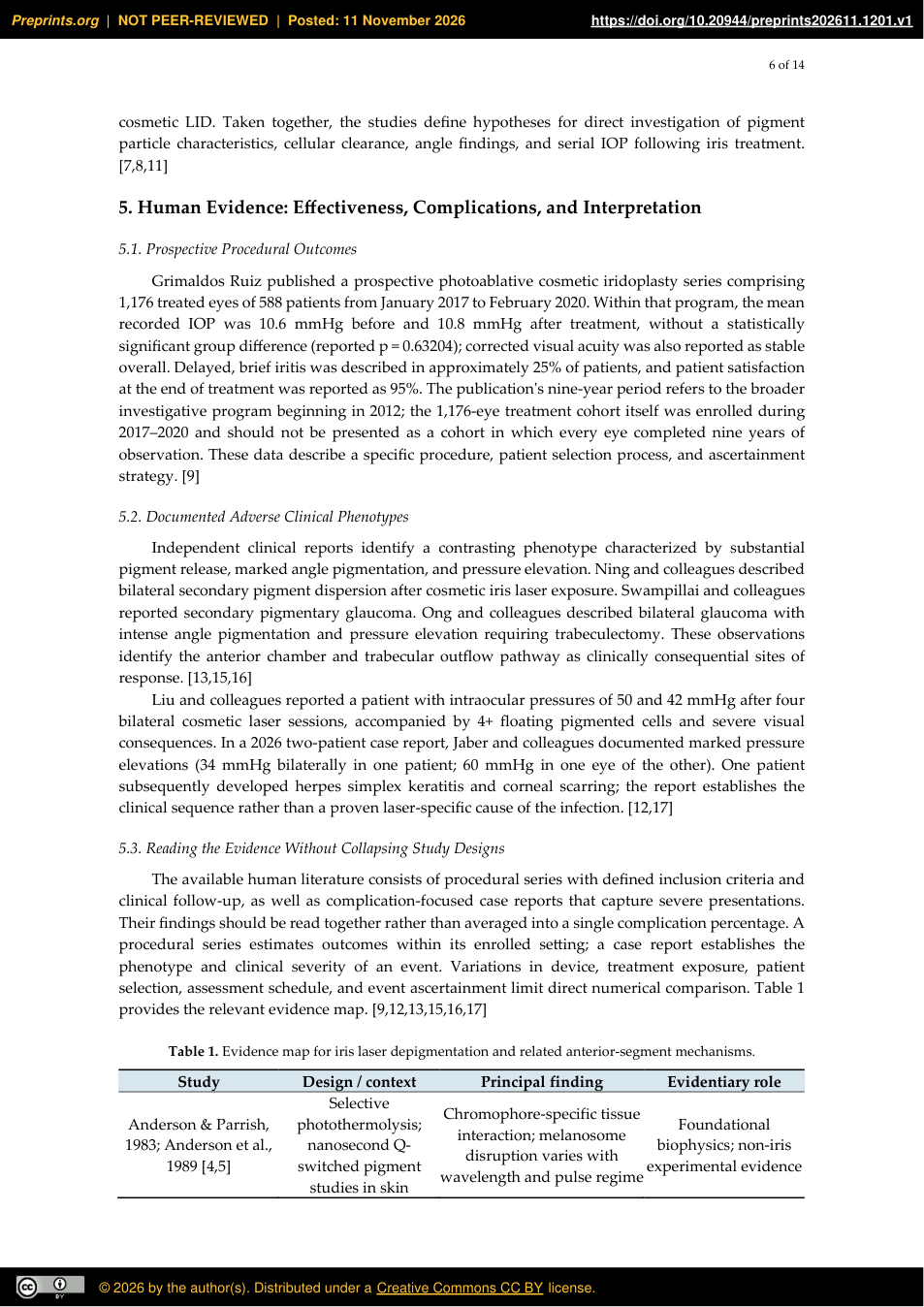
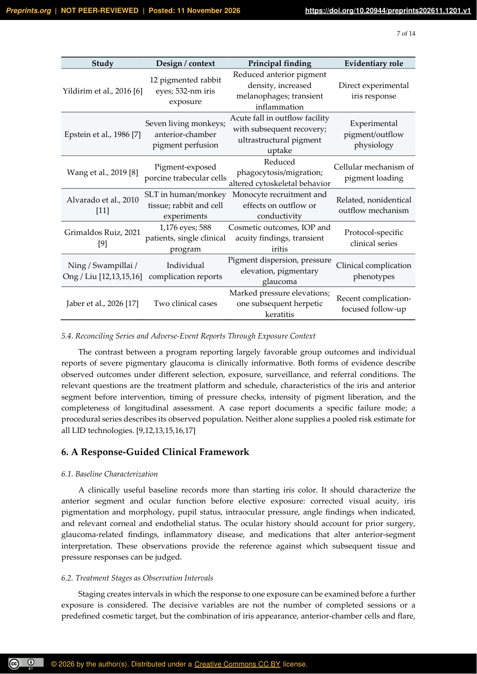
The available human literature consists of procedural series with defined inclusion criteria and clinical follow-up, as well as complication-focused case reports that capture severe presentations. Their findings should be read together rather than averaged into a single complication percentage. A procedural series estimates outcomes within its enrolled setting; a case report establishes the phenotype and clinical severity of an event. Variations in device, treatment exposure, patient selection, assessment schedule, and event ascertainment limit direct numerical comparison. Table 1 provides the relevant evidence map. [9,12,13,15,16,17]
Table 1. Evidence map for iris laser depigmentation and related anterior-segment mechanisms.
Study Design / context Principal finding Evidentiary role
Selective photothermolysis; nanosecond Qswitched pigment studies in skin
Chromophore-specific tissue interaction; melanosome disruption varies with wavelength and pulse regime
Anderson & Parrish, 1983; Anderson et al., 1989 [4,5]
Foundational biophysics; non-iris experimental evidence
Source page 7
Study Design / context Principal finding Evidentiary role
Reduced anterior pigment density, increased melanophages; transient inflammation
Yildirim et al., 2016 [6] 12 pigmented rabbit eyes; 532-nm iris exposure
Direct experimental iris response
Acute fall in outflow facility
Seven living monkeys;
Experimental pigment/outflow
with subsequent recovery;
Epstein et al., 1986 [7]
anterior-chamber pigment perfusion
ultrastructural pigment
physiology
uptake
Reduced phagocytosis/migration; altered cytoskeletal behavior
Wang et al., 2019 [8] Pigment-exposed porcine trabecular cells
Cellular mechanism of pigment loading
SLT in human/monkey
Monocyte recruitment and
Alvarado et al., 2010
Related, nonidentical
tissue; rabbit and cell
effects on outflow or
[11]
outflow mechanism
experiments
conductivity
1,176 eyes; 588 patients, single clinical program
Cosmetic outcomes, IOP and acuity findings, transient iritis
Grimaldos Ruiz, 2021 [9]
Protocol-specific clinical series
Pigment dispersion, pressure
Ning / Swampillai / Ong / Liu [12,13,15,16]
Individual complication reports
Clinical complication
elevation, pigmentary
phenotypes
glaucoma
Jaber et al., 2026 [17] Two clinical cases Marked pressure elevations; one subsequent herpetic keratitis
Recent complicationfocused follow-up
5.4. Reconciling Series and Adverse-Event Reports Through Exposure Context
The contrast between a program reporting largely favorable group outcomes and individual reports of severe pigmentary glaucoma is clinically informative. Both forms of evidence describe observed outcomes under different selection, exposure, surveillance, and referral conditions. The relevant questions are the treatment platform and schedule, characteristics of the iris and anterior segment before intervention, timing of pressure checks, intensity of pigment liberation, and the completeness of longitudinal assessment. A case report documents a specific failure mode; a procedural series describes its observed population. Neither alone supplies a pooled risk estimate for all LID technologies. [9,12,13,15,16,17]
6. A Response-Guided Clinical Framework
6.1. Baseline Characterization
A clinically useful baseline records more than starting iris color. It should characterize the anterior segment and ocular function before elective exposure: corrected visual acuity, iris pigmentation and morphology, pupil status, intraocular pressure, angle findings when indicated, and relevant corneal and endothelial status. The ocular history should account for prior surgery, glaucoma-related findings, inflammatory disease, and medications that alter anterior-segment interpretation. These observations provide the reference against which subsequent tissue and pressure responses can be judged.
6.2. Treatment Stages as Observation Intervals
Staging creates intervals in which the response to one exposure can be examined before a further exposure is considered. The decisive variables are not the number of completed sessions or a predefined cosmetic target, but the combination of iris appearance, anterior-chamber cells and flare,
Source page 8
pressure trajectory, angle findings, symptoms, and tissue stability. A stable visible color and normal ocular physiology represent separate components of the treatment assessment. [9,12,17] The selective stromal melanin modulation (SSMM) framework distinguishes biophysical, anatomical, and functional aspects of selectivity, emphasizing that pigment interaction, tissue preservation, and clinical response are separable domains. The related L-SAFE proposal organizes pre-exposure assessment and reassessment using ocular pressure, inflammation, corneal condition, and angle/outflow findings. In this review these constructs serve as a structured vocabulary for response-guided clinical decisions; they are not presented as independently validated prediction tools. [18,19]
6.3. Monitoring Tied to the Expected Biological Sequence
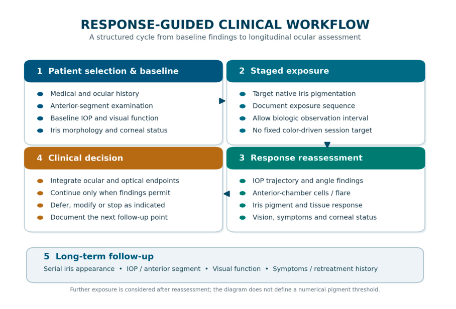
Early follow-up addresses acute pressure response, symptoms, anterior-chamber reaction, and obvious pigment dispersion. Subsequent assessments document resolution or persistence of these findings, evolution of visible iris color, and emerging corneal, angle, or optic-nerve abnormalities. Longer-term follow-up evaluates the stability of both appearance and ocular function. The schedule should be adapted to findings rather than treated as a fixed number of visits. An unexpected pressure increase, persistent cells or flare, symptomatic visual change, or progression of structural findings should trigger reassessment of further exposure and management of the ocular condition. Figure 2 expresses the clinical cycle as a sequence of observable checkpoints rather than a claim that any particular exposure schedule prevents complications. [12,13,17,19]
Figure 2. Response-guided clinical workflow. Baseline evaluation, staged exposure, reassessment, treatment
decision, and long-term follow-up are recurring components of clinical management. The workflow is a
proposed clinical decision structure, not a validated numerical decision rule. [9,18,19].
6.4. Suitability, Expectations, and Procedural Endpoints
Because elective color modification is not a treatment for visual disease, the justification for repeated exposure depends on both the patientʹs informed objectives and the eyeʹs observed response. A documented baseline, clear explanation of possible partial or heterogeneous color change, realistic description of further stages, and an explicit plan for reassessment support coherent clinical decisions. The extent of acceptable treatment is defined by ocular findings as well as cosmetic
Source page 9
goals; stopping the sequence remains a legitimate clinical endpoint when additional exposure is unwarranted. Meaningful follow-up records at least three types of outcome: visible iris change, patientexperienced visual symptoms, and objective ocular function. Photophobia, glare, changes in visual acuity, and inflammatory symptoms deserve documentation alongside pressure and slit-lamp findings. A stable photographic result and an otherwise uncomplicated clinical course are not synonymous; both should be assessed on their own terms. [9,12,17]
6.5. Selection, Monitoring, and Endothelial Documentation
Selection begins with the anatomical state of the eye. Anterior chamber configuration, a history of glaucoma or ocular inflammation, prior ocular surgery, pupillary or iris abnormalities, and existing corneal disease can materially change the interpretation of post-exposure findings. Baseline corrected vision, pupil assessment, slit-lamp findings, IOP, and anterior-segment imaging when clinically indicated provide the reference against which later responses can be judged. [10,12,13,17] Corneal endothelial assessment is a complementary structural endpoint. It is particularly relevant when repeated anterior-segment interventions are contemplated or the baseline endothelium is abnormal. A specular-microscopy measurement can document endothelial cell density and morphological characteristics at a defined observation point; serial results should be interpreted in the context of measurement variability and other ocular exposures. The endothelium is not the target of LID, but a complete longitudinal account of anterior-segment status should not rely on IOP alone. [3,20] The treatment record should preserve the sequence of exposures and the interval-specific findings rather than only the starting and final color photographs. This creates an auditable link between each treatment stage and the decision made afterward: continue, defer, modify, or conclude the course. Such documentation permits the same eye to be evaluated both as an optical outcome and as a longitudinal clinical record. [9,18,19]
7. Color Evolution, Optical Variability, and Long-Term Stability
7.1. Pigment Removal Does Not Uniquely Determine Hue
Visible iris appearance depends on pigment absorption and light propagation through the remaining stroma. Two irides with similar apparent degrees of pigment loss may differ in hue or brightness because of differences in pigment depth, residual distribution, stromal organization, and viewing conditions. This is consistent with established morphologic correlates of iris color and the range of outcomes reported in clinical photoablative iridoplasty. [1,2,9] For longitudinal evaluation, standardized photographs should be obtained with reproducible illumination, camera geometry, pupil size when feasible, exposure, and white balance. Such documentation quantifies appearance more reliably than non-standardized photographs but does not measure the absolute amount of melanin or establish stromal ultrastructure. When available, anterior-segment imaging and clinical examination provide complementary anatomical information.
7.2. Three Distinct Longitudinal Endpoints
Three endpoints should be recorded separately: (1) the change in visible iris appearance relative to baseline; (2) stability of that appearance within a defined follow-up interval; and (3) ocular functional and structural status during the same interval. A persistent color result is evidence of observed stability. Biological permanence is a different claim because finite observation cannot determine the full future course of pigment biology. Published follow-up is most useful when its duration, missing data, and assessment methods are specified. [9]
Source page 10
7.3. Repigmentation and Delayed Change
Potential sources of delayed appearance change include redistribution of residual pigment, evolving stromal optical conditions, and biological changes in pigment-bearing cells. Existing LID studies do not provide a standardized human longitudinal measurement of pigment content that would distinguish all such mechanisms. A sound follow-up design therefore records changes in appearance without automatically assigning them to melanocyte regeneration or irreversible pigment depletion. [1,9]
7.4. Cumulative Exposure and the Meaning of a Final Result
Repeated treatment introduces a second time dimension beyond the response to an individual session: the sequence of previous exposures and the condition of the eye at each return visit. The observed final appearance can therefore be described by its baseline state, number and timing of exposures, and documented follow-up period. Such a record is more informative than an unqualified designation of a color as permanent. It also enables comparison of eyes with similar photographic results but different treatment histories and physiological responses. [9]
7.5. Stability, Pigmentation, and the Meaning of Permanence
Three statements must remain distinct. Pigment reduction is a biological alteration documented directly in experimental iris histology; sustained lightening is an observed optical outcome over a defined follow-up interval; biological permanence would imply future behavior beyond the observation window. A stable photograph cannot distinguish all possible contributions of residual pigment, stromal scattering, changing illumination, or delayed biological adaptation. The evidentiary standard for long-term color stability is therefore serial, standardized observation with explicit time points, not a one-time photograph or an indefinite label. [1,2,6,9] Reports of limited long-term color change should be expressed as findings within their measured follow-up. A protocol-specific record of persistent appearance is useful clinical evidence even without a claim that every iris will respond identically or remain unchanged indefinitely. Tracking retreatment, inter-eye differences, changes in brightness or hue, and ocular function in parallel would help separate durability of the visible result from the condition of the treated tissue. [9]
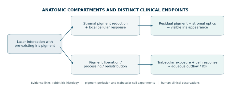
Figure 3. Two linked pathways following iris laser exposure. Pigment-related stromal optical change influences
visible appearance, whereas released pigment and cellular responses can influence the anterior chamber and
aqueous outflow. The two domains are clinically connected but are evaluated through different measurements.
[6,7,8,11].
Source page 11
8. Anatomical Comparison With Other Eye-Color Procedures
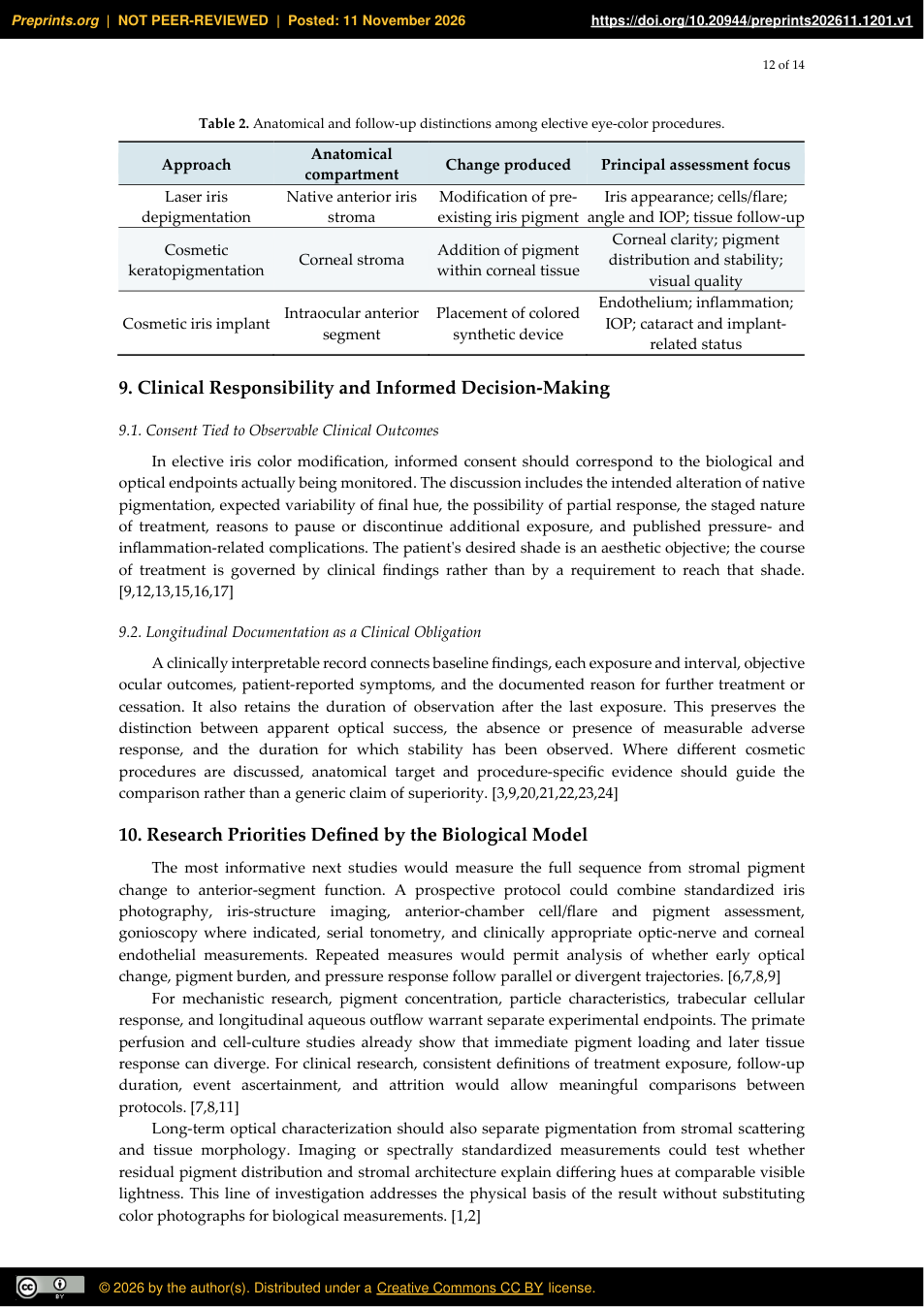
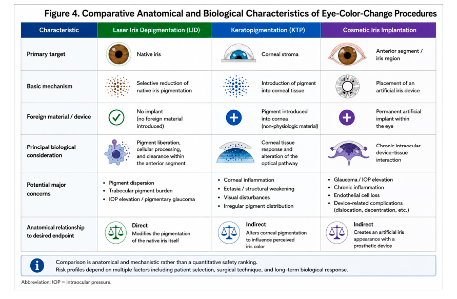
Laser iris depigmentation, cosmetic keratopigmentation, and cosmetic iris implantation should be compared according to the anatomical structure changed, the material introduced or removed, and the clinically relevant follow-up endpoints. LID modifies native iris pigment. Keratopigmentation introduces colorant into corneal tissue, including femtosecond-assisted intrastromal approaches. Cosmetic iris implantation places an artificial colored device inside the anterior segment. This anatomical distinction determines the type of tissue interaction under evaluation. [3,20] Keratopigmentation evidence includes technique-specific clinical series and reviews distinguishing purely cosmetic procedures from therapeutic applications in eyes with pre-existing pathology. A prospective single-surgeon series of 166 eyes treated for purely cosmetic indications found no statistically significant mean change in visual acuity, visual-field mean deviation, or IOP at 12 months. The broader literature documents postoperative light sensitivity, pigment redistribution or color change, and, in selected cosmetic reports, corneal ectatic change. These observations concern a different tissue compartment and should be interpreted according to indication, procedure, followup interval, and study design. [21,22] Cosmetic iris implants have a substantially different mechanism and complication profile, with reported corneal decompensation, glaucoma, uveitis, cataract, and need for explantation or additional surgery. A French multicenter retrospective series included 87 reported eyes, with 65 eyes having complete questionnaires for analysis; a later tertiary-center series described 30 eyes of 15 patients treated for implant complications. These referral-based cohorts document serious clinical events in affected patients and should be interpreted in their specific ascertainment settings. [23,24] Direct comparisons should retain these anatomical and methodological distinctions. Table 2 summarizes the procedure, altered compartment, and principal follow-up measurements; it is intended for clinical interpretation rather than assigning a single numerical rank to different interventions. [3,20]
Figure 4. Comparative anatomical and biological characteristics of laser iris depigmentation, cosmetic
keratopigmentation, and cosmetic iris implantation. The figure summarizes anatomical targets, introduced
material and documented considerations; it does not rank procedures by comparative safety. [3,20,21,22,23,24].
Source page 12
Table 2. Anatomical and follow-up distinctions among elective eye-color procedures.
Approach Anatomical compartment Change produced Principal assessment focus
Laser iris depigmentation Native anterior iris stroma Modification of preexisting iris pigment Iris appearance; cells/flare; angle and IOP; tissue follow-up
Corneal clarity; pigment distribution and stability;
Cosmetic keratopigmentation Corneal stroma Addition of pigment within corneal tissue
visual quality
Endothelium; inflammation; IOP; cataract and implantrelated status
Cosmetic iris implant Intraocular anterior segment Placement of colored synthetic device
9. Clinical Responsibility and Informed Decision-Making
9.1. Consent Tied to Observable Clinical Outcomes
In elective iris color modification, informed consent should correspond to the biological and optical endpoints actually being monitored. The discussion includes the intended alteration of native pigmentation, expected variability of final hue, the possibility of partial response, the staged nature of treatment, reasons to pause or discontinue additional exposure, and published pressure- and inflammation-related complications. The patientʹs desired shade is an aesthetic objective; the course of treatment is governed by clinical findings rather than by a requirement to reach that shade. [9,12,13,15,16,17]
9.2. Longitudinal Documentation as a Clinical Obligation
A clinically interpretable record connects baseline findings, each exposure and interval, objective ocular outcomes, patient-reported symptoms, and the documented reason for further treatment or cessation. It also retains the duration of observation after the last exposure. This preserves the distinction between apparent optical success, the absence or presence of measurable adverse response, and the duration for which stability has been observed. Where different cosmetic procedures are discussed, anatomical target and procedure-specific evidence should guide the comparison rather than a generic claim of superiority. [3,9,20,21,22,23,24]
10. Research Priorities Defined by the Biological Model
The most informative next studies would measure the full sequence from stromal pigment change to anterior-segment function. A prospective protocol could combine standardized iris photography, iris-structure imaging, anterior-chamber cell/flare and pigment assessment, gonioscopy where indicated, serial tonometry, and clinically appropriate optic-nerve and corneal endothelial measurements. Repeated measures would permit analysis of whether early optical change, pigment burden, and pressure response follow parallel or divergent trajectories. [6,7,8,9] For mechanistic research, pigment concentration, particle characteristics, trabecular cellular response, and longitudinal aqueous outflow warrant separate experimental endpoints. The primate perfusion and cell-culture studies already show that immediate pigment loading and later tissue response can diverge. For clinical research, consistent definitions of treatment exposure, follow-up duration, event ascertainment, and attrition would allow meaningful comparisons between protocols. [7,8,11] Long-term optical characterization should also separate pigmentation from stromal scattering and tissue morphology. Imaging or spectrally standardized measurements could test whether residual pigment distribution and stromal architecture explain differing hues at comparable visible lightness. This line of investigation addresses the physical basis of the result without substituting color photographs for biological measurements. [1,2]
Source page 13
10.1. Minimum Common Dataset for Comparative Studies
A shared reporting dataset would improve interpretation across centers: number of patients and eyes, baseline pigmentation and ocular eligibility, description of the laser platform and exposure schedule, follow-up distribution, retreatment frequency, serial IOP, inflammatory and pigment findings, vision-related outcomes, endothelial measurements where obtained, and explicit definitions of adverse events. At each follow-up interval, investigators should distinguish the eyes evaluated from those originally treated. A cohort with a clear denominator and longitudinal completeness can answer incidence and stability questions that isolated complications or heterogeneous narratives cannot. [9,12,17]
11. Conclusion
Laser iris depigmentation acts on native iris pigmentation within a living optical and anatomical system. Experimental iris histology demonstrates pigment modification and cellular processing; pigment-perfusion and trabecular-cell studies establish biological pathways through which released material can influence outflow tissue; human series and complication reports describe the range of observable clinical responses. Together, these findings support a model in which the visible response and anterior-segment physiological response are related, distinct, and jointly relevant to staged treatment. [6,7,8,9,12,13,15,16,17] Controlled pigment dispersion provides a longitudinal description of treatment-related pigment liberation and handling, while the response-guided clinical framework specifies how that response is observed between exposures. Clinical endpoints encompass iris appearance, tissue status, angle findings, IOP, inflammation, symptoms, and visual function. Separating pigment reduction, observed long-term stability, and biological permanence allows outcomes to be reported precisely. The resulting synthesis connects experimental mechanisms to clinical documentation and defines measurable priorities for future comparative research. [1,2,6,7,8,9,18,19]
References
1. Imesch PD, Wallow IHL, Albert DM. The color of the human eye: a review of morphologic correlates and
of some conditions that affect iridial pigmentation. Surv Ophthalmol. 1997;41(Suppl 2):S117–S123.
doi:10.1016/S0039-6257(97)80018-5.
2. Sturm RA, Larsson M. Genetics of human iris colour and patterns. Pigment Cell Melanoma Res.
2009;22:544–562. doi:10.1111/j.1755-148X.2009.00606.x.
3. D’Oria F, Abu-Mustafa SK, Alio JL. Cosmetic change of the apparent color of the eye: a review on surgical
alternatives, outcomes and complications. Ophthalmol Ther. 2022;11:465–477. doi:10.1007/s40123-022-
00458-2.
4. Anderson RR, Parrish JA. Selective photothermolysis: precise microsurgery by selective absorption of
pulsed radiation. Science. 1983;220(4596):524–527. doi:10.1126/science.6836297.
5. Anderson RR, Margolis RJ, Watenabe S, Flotte T, Hruza GJ, Dover JS. Selective photothermolysis of
cutaneous pigmentation by Q-switched Nd:YAG laser pulses at 1064, 532, and 355 nm. J Invest Dermatol.
1989;93(1):28–32. doi:10.1111/1523-1747.ep12277339.
6. Yildirim Y, Duzgun E, Kar T, Sonmez M, Kucukodaci Z, Ersanli D, et al. Evaluation of color-changing effect
and complications after Nd:YAG laser application on iris surface. Med Sci Monit. 2016;22:107–114.
doi:10.12659/MSM.895086.
7. Epstein DL, Freddo TF, Anderson PJ, Patterson MM, Bassett-Chu S. Experimental obstruction to aqueous
outflow by pigment particles in living monkeys. Invest Ophthalmol Vis Sci. 1986;27:387–395.
PMID:3949467.
8. Wang C, Dang Y, Loewen RT, Waxman S, Shah P, Xia X, et al. Impact of pigment dispersion on trabecular
meshwork cells. Graefes Arch Clin Exp Ophthalmol. 2019;257:1217–1230. doi:10.1007/s00417-019-04300-7.
9. Grimaldos Ruiz P. Photoablative cosmetic iridoplasty: effective, safe, and predictable—eye color change in
1176 eyes. Int Ophthalmol. 2021;41:1381–1393. doi:10.1007/s10792-021-01693-5.
Source page 14
10. Scuderi G, Contestabile MT, Scuderi L, Librando A, Fenicia V, Rahimi S. Pigment dispersion syndrome and
pigmentary glaucoma: a review and update. Int Ophthalmol. 2019;39:1651–1662. doi:10.1007/s10792-018-
0938-7.
11. Alvarado JA, Katz LJ, Trivedi S, Shifera AS. Monocyte modulation of aqueous outflow and recruitment to
the trabecular meshwork following selective laser trabeculoplasty. Arch Ophthalmol. 2010;128:731–737.
doi:10.1001/archophthalmol.2010.85.
12. Liu J, Korban S, Moster MR, Rhéaume MA, Wang Q. Bilateral severe iatrogenic pigmentary glaucoma
following laser treatment for cosmetic iris color change. Am J Ophthalmol Case Rep. 2023;32:101927.
doi:10.1016/j.ajoc.2023.101927.
13. Ong AY, Ching-A-Sue G, Krilis M, Guirao-Navarro MC, Hornby S, Jutley G. Refractory iatrogenic
pigmentary glaucoma secondary to cosmetic laser treatment: a case report. Eur J Ophthalmol. 2023;33:NP1–
NP4. doi:10.1177/11206721211050338.
14. Mete M. Temporal Response Architecture After Laser-Based Iris Pigment Modification: A Mechanistic
Synthesis of Anterior-Segment Response Domains. Version 1.1. Zenodo [preprint]. 2026.
doi:10.5281/zenodo.23128049.
15. Ning B, Baboolal S, Gizzi C, Nolan W. A case of secondary pigment dispersion following laser to
cosmetically lighten the irises. J Glaucoma. 2022;31:133–135. doi:10.1097/IJG.0000000000001790.
16. Swampillai AJ, Sherman T, Garg A, Tan IJ, Lim KS. Secondary pigmentary glaucoma following cosmetic
laser treatment to alter iris colour. Cont Lens Anterior Eye. 2023;46:101754. doi:10.1016/j.clae.2022.101754.
17. Jaber W, Nemet M, Waisbourd M. Laser iris depigmentation resulting in markedly elevated intraocular
pressure and herpes simplex keratitis with corneal scarring: case report. Case Rep Ophthalmol.
2026;17:312–317. doi:10.1159/000551226.
18. Mete M. Selective Stromal Melanin Modulation: A Three-Level Framework for Evaluating Biophysical,
Anatomical, and Functional Selectivity. Zenodo. 2026. doi:10.5281/zenodo.23424601.
19. Mete M. Lumineyes Anterior-Segment Safety Staging (L-SAFE): A Proposed Framework for Pre-Exposure
Assessment in Laser Iris Pigment Reduction. Zenodo. 2026. doi:10.5281/zenodo.23046455.
20. D’Oria F, Alio JL. Surgical techniques for cosmetic eye color change: a narrative review. Ophthalmol Ther.
2025;14:1685–1694. doi:10.1007/s40123-025-01177-0.
21. Alio JL, Sanginabadi A, Hojabr AT, Jafari B. Femtosecond laser-assisted keratopigmentation outcomes for
pure cosmetic purposes. Am J Ophthalmol Case Rep. 2025;38:102297. doi:10.1016/j.ajoc.2025.102297.
22. Hashemi H, Hadavandkhani A, Khabazkhoob M. An updated comprehensive review on
keratopigmentation. Int J Ophthalmol. 2025;18:2183–2194. doi:10.18240/ijo.2025.11.22.
23. El Chehab H, Gatinel D, Baudouin C, Muraine M, Hoffart L, Rozot P, et al. Complications of cosmetic iris
implants: French series of 87 eyes. J Cataract Refract Surg. 2020;46:34–39.
doi:10.1097/j.jcrs.0000000000000032.
24. Balal S, Jamall O, Janmohamed IK, Hossain I, Hussain R, Kim SE, et al. Complications and management of
cosmetic iris implants. Am J Ophthalmol. 2025;277:433–440. doi:10.1016/j.ajo.2025.05.049.
of the individual author(s) and contributor(s) and not of MDPI and/or the editor(s). MDPI and/or the editor(s)
disclaim responsibility for any injury to people or property resulting from any ideas, methods, instructions or
products referred to in the content.
Original table layouts
Images below preserve the typeset table layouts in the source PDF. The full published PDF remains the authoritative layout reference.