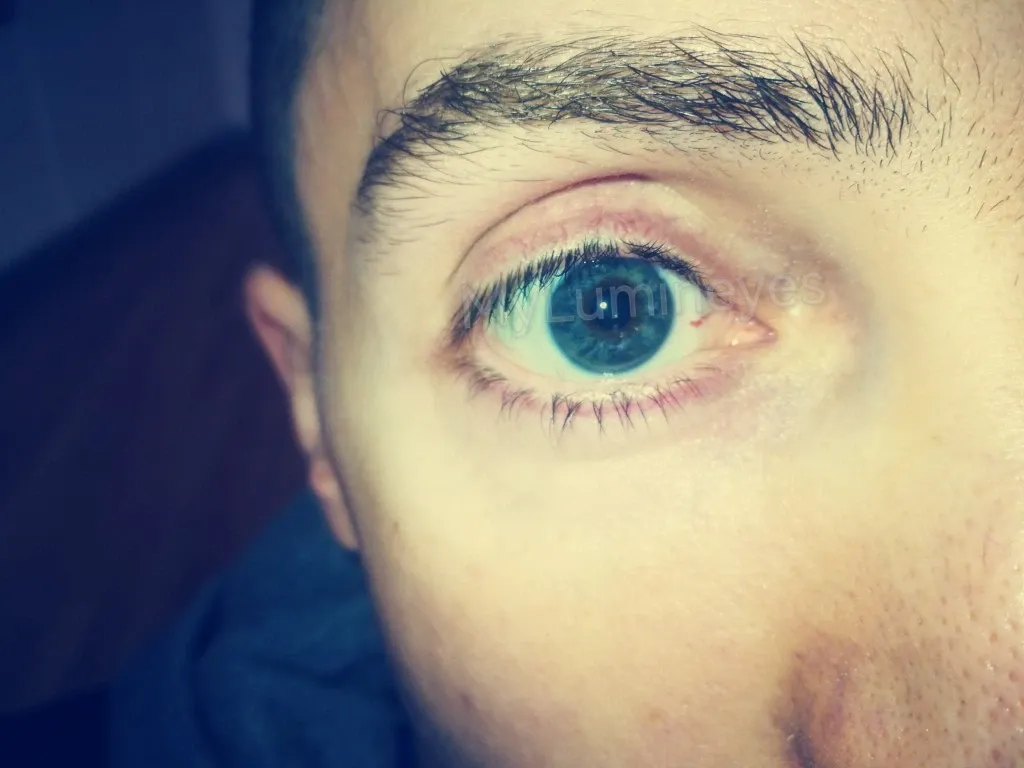
Heterochromia refers to a difference in color between the two eyes or within different areas of the same iris. It is an uncommon but usually harmless condition that occurs when melanin distribution varies between the irises. Most cases are congenital, while others may appear later in life due to medical or structural changes.
The appearance of heterochromia depends on melanin levels within the iris stroma. When one eye contains more pigment than the other, the colors differ. This variation does not typically affect vision or eye health.
Types of Heterochromia
Heterochromia is classified into three forms:
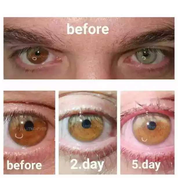
Complete heterochromia: Each eye has a distinctly different color (for example, one blue and one brown).
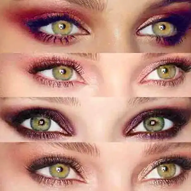
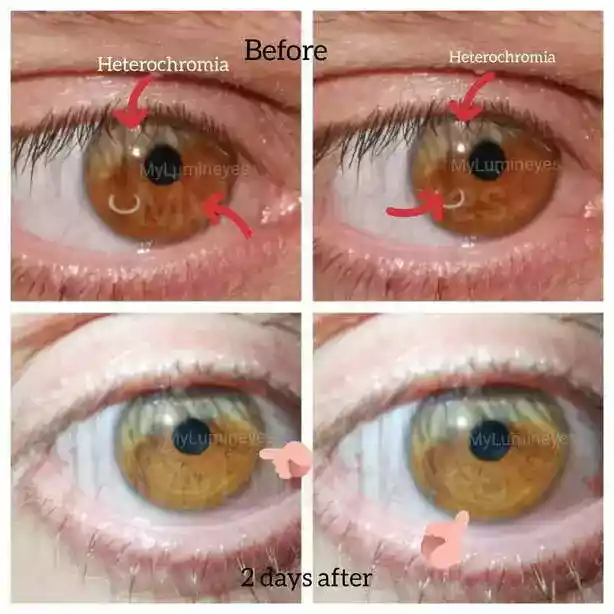
Sectoral heterochromia: A segment of one iris differs in color from the rest of the same eye.
Central heterochromia: The inner ring around the pupil shows a different color compared to the outer iris.
What Causes Heterochromia?
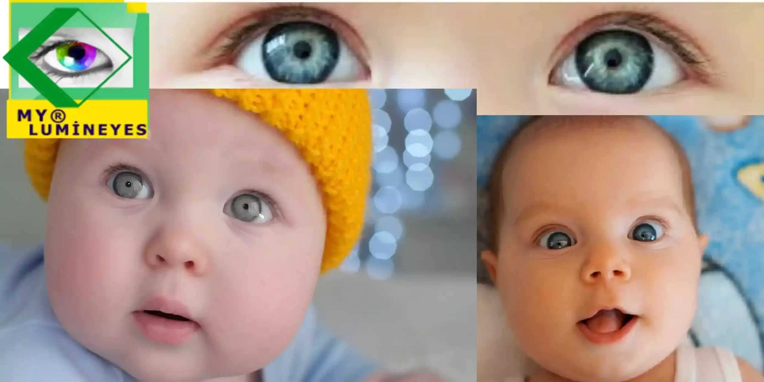
Most congenital cases arise from genetic variations affecting melanin distribution. These variations occur during early development and are typically stable throughout life.
Acquired heterochromia may occur due to:
• Trauma
• Inflammation
• Certain medications (such as prostaglandin analogues)
• Structural iris changes
• Pigment dispersion or deposition
A medical examination is recommended when heterochromia appears suddenly or is associated with pain, redness or vision changes.
Does Heterochromia Affect Vision?
In the majority of cases, heterochromia does not impact visual function. The variation is purely cosmetic. However, when caused by ocular disease or previous trauma, vision may be affected depending on the underlying condition.
Can Heterochromia Change Over Time?
Congenital heterochromia usually remains stable. Acquired cases may evolve based on pigment activity, inflammation or medication exposure. Prostaglandin-based drops used in glaucoma treatment may increase melanin production, occasionally deepening iris color.
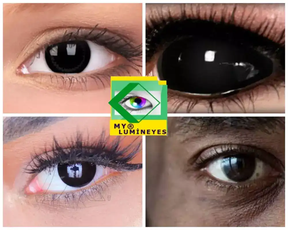
Is There a Way to Correct or Equalize Eye Colors?
Topical or nutritional methods cannot influence melanin in the iris. Color contact lenses are the safest temporary solution for cosmetic symmetry.
In selected medical cases, controlled procedures that interact with iris melanin may be evaluated by specialized centers. These approaches aim to achieve color balance without affecting ocular structures. Detailed information on pigment-targeting methods can be found here: Laser Eye Color Change.
When Should Someone See a Doctor?
A professional evaluation is recommended if heterochromia develops suddenly, progresses quickly or is accompanied by ocular symptoms. Early examination helps identify treatable conditions and ensures long-term eye health.
Conclusion
Heterochromia is a rare and visually distinctive condition that results from differences in iris melanin. While most cases are benign and stable, acquired forms may require medical assessment. Understanding its causes and structure helps distinguish natural variations from underlying ocular conditions.